Method for establishing cavernous body fibrosis disease model

A technology for fibrotic diseases and model establishment, which is applied in the fields of biochemical equipment and methods, pharmaceutical formulations, and preparations for in vivo tests, etc., to achieve the effects of convenient administration, easy dosage control, and short modeling time.

Active Publication Date: 2022-07-29
SHANGHAI FIRST PEOPLES HOSPITAL
View PDF7 Cites 0 Cited by
  • Summary
  • Abstract
  • Description
  • Claims
  • Application Information

AI Technical Summary

Problems solved by technology

[0006] However, there are no reports on in vitro disease models and animal models that simulate endothelial-mesenchymal cell transformation leading to fibrosis of cavernous tissue.

Method used


Image

  • Method for establishing cavernous body fibrosis disease model
  • Method for establishing cavernous body fibrosis disease model

Examples


Embodiment 1

[0024] Materials and Methods

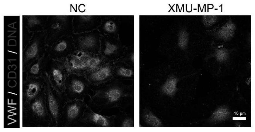
[0025] 1. Human cavernous endothelial cell isolation and XMU-MP-1 treatment

[0026] A fresh corpus cavernosum tissue sample, approximately 1 x 1 cm in size, was first immersed in 15 ml of PBS and shaken vigorously to remove residual blood. The tissue was cut into 1×1 mm pieces and digested with 2.5 mg / ml collagenase type I, 4 mg / ml collagenase IV, 0.1 mg / ml neutral protease and 2 mg / ml DNase I enzyme for 20 min at 37°C. Subsequently, the cell suspension was filtered through a 40 mm nylon mesh, and cells were sorted by MACS and a dead cell removal kit (Miltenyi Biotec) to remove dead cells. The digested cell suspension was washed and resuspended in EGM-2 (CC-3202; Lonza) medium. Cell colonies began to appear around day 5 of culture, followed by the addition of fibroblast inhibitor for 7-10 days. When the isolated cell clones began to confluent with each other, the ENC clones were labeled and digested using a cloning cylinder (C7983-50EA; 809Si...


PUM


Abstract

The invention relates to a method for establishing a cavernous body fibrosis disease model. The method comprises the following steps: (1) separating endothelial cells of a cavernous body and carrying out XMU-MP-1 treatment on the endothelial cells of the cavernous body; (2) carrying out immunofluorescence staining on the cell slide; (3) carrying out in-vitro culture and XMU-MP-1 treatment on the cavernous body tissue; (4) performing Masson dyeing on the cavernous body tissue paraffin section; and (5) carrying out XMU-MP-1 treatment on the in-vivo cavernous body of the rat, so as to obtain the required endothelial-mesenchymal transition mediated cavernous body fibrosis rat model. According to the invention, the blank of lack of a cavernous body fibrosis model caused by endothelial-mesenchymal transition at present is filled, and the development of in-vivo and in-vitro experiments of the phenomenon is greatly facilitated. The XMU-MP-1 used in the invention is used for activating a Hippo pathway of cavernous body fibroblasts, has small influence on other biological processes causing cavernous body fibrosis, such as fibroblast-myofibroblast conversion, fibroblast proliferation and the like, and has target specificity. Therefore, aiming at the biological process of endothelial-mesenchymal transformation, the interference can be eliminated by using the method for modeling.

Description

technical field [0001] The invention relates to the technical field of animal model establishment, in particular to a method for establishing a cavernosal fibrosis disease model. Background technique [0002] In the field of ED research, there is no suitable in vitro disease model and animal model that simulates endothelial-mesenchymal transition leading to fibrosis in cavernous tissue. At present, the commonly used models of fibrosis of cavernous tissue induced by other ways include TGFβ-induced model, diabetic rat model and aging rat model. [0003] The above-mentioned models take a long time to build, and at the same time, they will lead to endothelial cell damage in the cavernosa and smooth muscle fibrosis, which leads to the introduction of multivariate interference in the experiment, which is not conducive to the experimental research on the process of endothelial-mesenchymal cell transition. In addition, the model establishment time is long. Elderly rats usually take...

Claims


Application Information

Patent Timeline
Patent Type & Authority Applications(China)
IPC IPC(8): C12N5/077A01K67/027C12Q1/02A61K49/00
CPCC12N5/0656A01K67/027G01N33/5008A61K49/0008A01K2207/20A01K2227/105A01K2267/03C12N2501/999C12N2503/02G01N2500/10Y02A50/30
Inventor 韩厦赵亮宇李铮姚晨成纪智勇罗嘉强李朋许俊伟徐帅
Owner SHANGHAI FIRST PEOPLES HOSPITAL
Why Eureka
Try Eureka