Region segmentation method and device

A region segmentation and image technology, applied in the field of medical image processing, can solve the problems of long segmentation time, inability to segment the ischemic core region and penumbra, limitations of images and data materials, etc., to achieve refined segmentation and shorten segmentation time. , Improve the effect of segmentation efficiency

Pending Publication Date: 2022-08-05
TONGXIN YILIAN TECH BEIJING
View PDF8 Cites 0 Cited by
  • Summary
  • Abstract
  • Description
  • Claims
  • Application Information

AI Technical Summary

Problems solved by technology

[0006] Embodiments of the present invention provide a region segmentation method, device, computer equipment, and storage medium to solve the problems in the related ar

Method used


Image

  • Region segmentation method and device
  • Region segmentation method and device
  • Region segmentation method and device

Examples


Embodiment 1

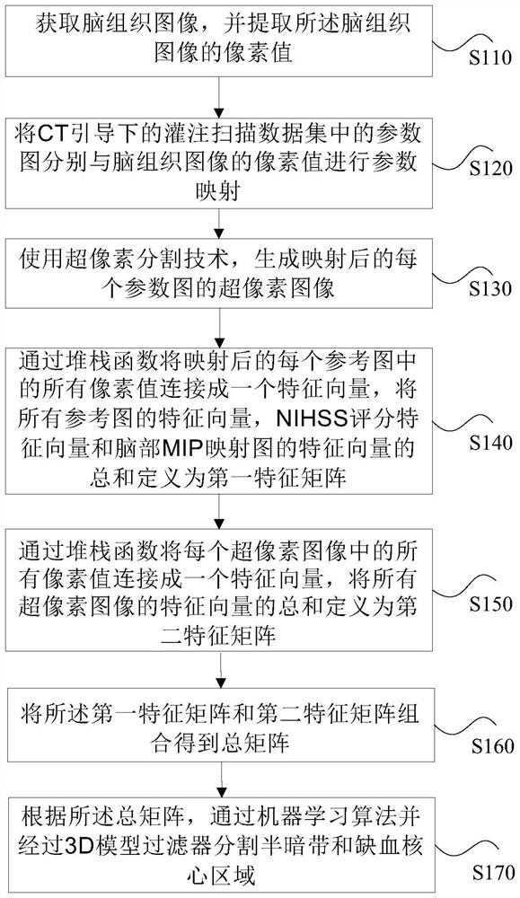
[0081] The present invention provides a regional segmentation method (single step method), such as figure 1 As well as 2 The flow chart shown, including:

[0082] Step S110, get brain tissue images, and extract the pixel value of the brain tissue image.

[0083] In this step, before dividing the ischemic area (semi -dark band and the core area of ​​ischemia), pre -processing needs to be performed, that is, extract brain tissue images from the brain image, and retain the use of brain tissue (BT) from the brain tissue (BT)) Pixel values.

[0084] Step S12, parameter maps of the parameter diagram of the scanning data concentration under the CT guidance, respectively, and the parameter map of the pixel value of the brain tissue image, which includes the reference map of the cerebral blood flow flow, the reference map of the cerebral blood capacity, the peak time reference diagram, the reference map, the reference Peak reference diagram, brain mip mapping diagram and NiHSS scoring tabl...

Embodiment 2

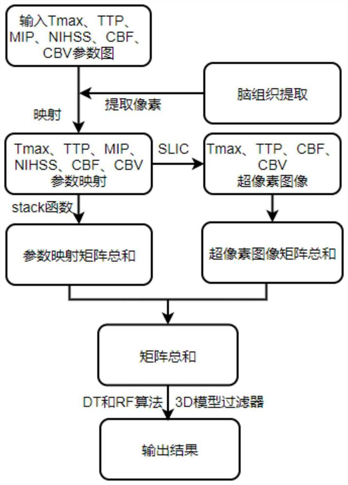
[0119] The embodiment of the present invention also provides another regional division method (two steps), such as Image 6 Show, including:

[0120] Step 1 (used to segment semi -dark belt):

[0121] Step s1.1, get brain tissue images, and extract the pixel value of the brain tissue image;

[0122] This step is the same as the step S110, so I do not repeat it here.

[0123] Step S1.2, the parameter diagram of the pouring scanned data concentration under CT guidance is performed with the pixel value of the brain tissue image, which includes the peak time reference map, the peak time reference map, the brain MIP mapping diagram And NiHSS scoring table;

[0124] In step S1.2, the peak time TTP reference map of the data concentration, the peak time TMAX reference map mapped to the extracted pixel value of the extracted brain tissue (BT) as the input feature. The specific mapping process is the same as S120. Research.

[0125] Step S1.3, use the ultra -pixel division technology to gene...

Embodiment 3

[0176] The embodiment of the present invention provides a regional segmentation device, such as Figure 10 Show, including:

[0177] The brain tissue obtains the module for obtaining brain tissue images, and extracts the pixel value of the brain tissue image;

[0178] The mapping module is used to perform parameter maps with the parameter diagram of the scanning data concentration under the CT guidance, respectively. , Dafeng Time Reference diagram, brain MIP mapping diagram and NiHSS scoring score table;

[0179] Ultra pixel image generation module is used to use ultra -pixel segmentation technology to generate the ultra -pixel image of each parameter diagram after mapping, including cerebral blood flow super pixel image, cerebral blood capacity ultra -pixel image, peak time ultra -pixel image and Dafeng peak Time super pixel image;

[0180] The first feature matrix generation module is used to connect all the pixel values ​​in each reference diagram into a feature vector through ...


PUM


Abstract

The invention discloses a region segmentation method and device. The method comprises the following steps: extracting a pixel value of a brain tissue image; performing parameter mapping on the parameter maps in the perfusion scanning data set and the pixel values of the brain tissue image; generating a superpixel image of each parameter graph by using a superpixel segmentation technology; defining the sum of the feature vectors of all the reference images, the NIHSS score feature vector and the feature vector of the brain MIP mapping image as a first feature matrix; defining the sum of the feature vectors of all the superpixel images as a second feature matrix; combining the first feature matrix and the second feature matrix to obtain a total matrix; and segmenting the penumbra and the ischemic core region through a machine learning algorithm and a 3D model filter. The full-automatic algorithm is adopted to segment the penumbra and the ischemic core area, and compared with a traditional segmentation technology, segmentation is more refined, segmentation time is greatly shortened, and segmentation efficiency is improved.

Description

Technical field [0001] This application involves the field of medical image processing technology. Specifically, it involves a regional segmentation method, device, computer equipment and storage media. Background technique [0002] For the segmentation of ischemic areas, it is generally used to "manually segmentation after computer fault scanning and irrigation technology". Through the computer fault scanning and irrigation (CTP), the three -dimensional (3D 3D (3D (3D (3D (3D (3D (3D (3D (3D (3D (3D (3D (3D (3D (3D (3D (3D (3D) during the injection of the contrast agent injection in CTP )data set. Based on the changes in the tissue density over time, the parameter chart of the color encoding is calculated, and these parameter diagrams are used to indirectly evaluate the ischemia core area and semi -band. Although manual segmentation technology is divided according to the patient's actual situation and adopted effective treatment decisions, the manual segmentation technology take...

Claims


Application Information

Patent Timeline
IPC IPC(8): G06T7/00G06T7/11G06V10/762G06V10/764G06K9/62G06N20/00
CPCG06T7/0012G06T7/11G06V10/762G06V10/764G06N20/00G06T2207/20081G06T2207/30016G06T2207/30104G06F18/2411G06F18/24323
Inventor 刘伟奇马学升陈金钢徐鹏赵友源庞盼陈磊
Owner TONGXIN YILIAN TECH BEIJING
Why Eureka
Try Eureka