Method and apparatus for performing intra-operative angiography

An angiography, vascular technology, used in surgery, blood flow measurement, medical science, etc.

Inactive Publication Date: 2003-02-26
NAT RES COUNCIL OF CANADA
View PDF0 Cites 37 Cited by
  • Summary
  • Abstract
  • Description
  • Claims
  • Application Information

AI Technical Summary

Problems solved by technology

Whilst the use of x-ray technology can assist physicians in determining whether a functioning fistula can be cre

Method used


Image

  • Method and apparatus for performing intra-operative angiography
  • Method and apparatus for performing intra-operative angiography

Examples


Embodiment Construction

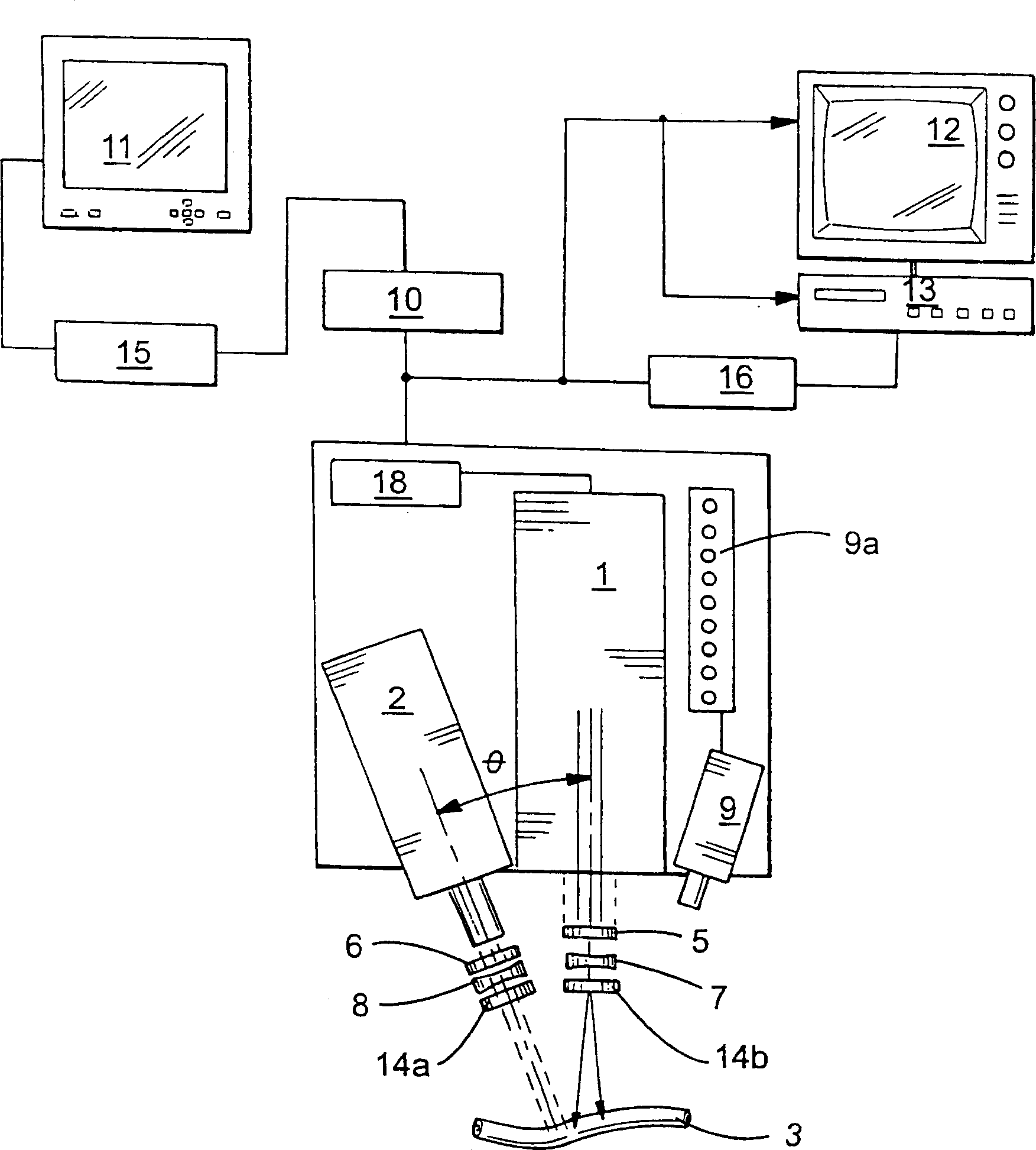
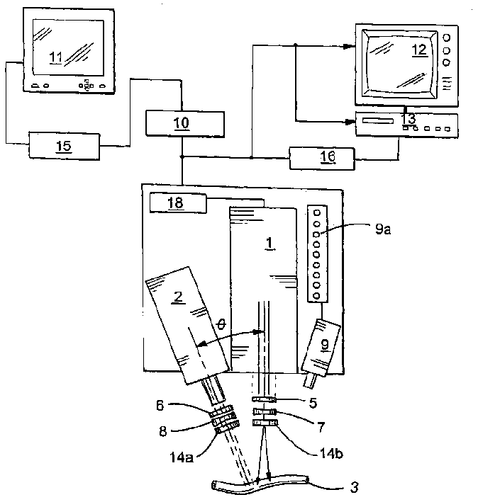
[0064] The described examples show the use of a preferred device of the present invention in observing the flow of fluorescent dye through a specific vessel, i.e. the mouse femoral artery, and the Langendorff perfused heart, and also show that the device works under normal conditions and under the influence of locally applied acetylcholine The ability to determine the diameter of mouse femoral artery vessels.

[0065] In this example, a fluorescent dye (ICG) is injected into the vascular bed (via a mouse jugular catheter: a perfusion catheter that perfuses the heart via a langendorff) and excited with the original radiation (806 nm) from a laser. Fluorescence (radiation) emitted by the dye (830 nm) is captured with a CCD camera as a series of vascular imaging images. The angiographic image relayed by the camera becomes the analog-to-digital conversion software running on the PC to digitize the angiographic image. These digitized images are then analyzed qualitatively (by view...


PUM


Abstract

The device is provided with a laser (1) for exciting the fluorescent imaging agent which emits radiation at a wavelength that causes any of the agent located within the vasculature or tissue of interest (3) irradiated thereby to emit radiation of a particular wavelength. Advantageously, a camera capable of obtaining multiple images over a period of time, such as a CCD camera (2) may be used to capture the emissions from the imaging agent. A band-pass filter (6) prevents the capture of radiation other than that emitted by the imaging agent. A distance sensor (9) incorporates a visual display (9a) providing feedback to the physician saying that the laser be located at a distance from the vessel of interest that is optimal for the capture of high quality images.

Description

technical field [0001] The present invention generally relates to methods for observing blood flow through the cardiovascular system of an animal. This patent application claims priority to US Patent Application No. 60 / 155.652, filed September 24,1999. Background of the invention [0002] Diseases and injuries affecting the cardiovascular system of animals, especially humans, are common in today's society. One such disease is atherosclerosis. The disease is characterized by partial blockage of blood vessels (vascular narrowing), typically narrowing of one or more arteries. In its most severe form, blood vessels are narrowed to complete blockage (atresia). In the coronary arteries, narrowing and atresia are usually most severe in the form of severe chest pain and, potentially, myocardial infarction (heart attack). Not limited to the coronary arteries, atherosclerosis can also affect the peripheral vasculature, that is, the arteries (and veins) that circulate blood in the ...

Claims


Application Information

Patent Timeline
IPC IPC(8): A61B5/00A61B1/00A61B5/026A61B5/0275A61B10/00
CPCA61B5/0275A61B5/0261
Inventor 约翰·C·多彻蒂马克·休考古尔普利特·曼加脱罗伯特·W·弗劳尔赛夏特里·M·查里
Owner NAT RES COUNCIL OF CANADA
Why Eureka
Try Eureka