System and method for measuring local lung function by CT using electronic beam

A lung function and detector technology, applied in the field of measuring local lung function, can solve problems such as inability to diagnose diseases, rough sampling, and patients receiving excessive radiation doses

Inactive Publication Date: 2004-07-14
GE MEDICAL SYST GLOBAL TECH CO LLC
View PDF12 Cites 9 Cited by
  • Summary
  • Abstract
  • Description
  • Claims
  • Application Information

AI Technical Summary

Problems solved by technology

Since the duration of the lung inhalation-exhalation action is only about two seconds, using 500 milliseconds would result in too coarse a sampling (low level of detail) of the patient's lungs to diagnose developing

Method used


Image

  • System and method for measuring local lung function by CT using electronic beam
  • System and method for measuring local lung function by CT using electronic beam
  • System and method for measuring local lung function by CT using electronic beam

Examples


Embodiment Construction

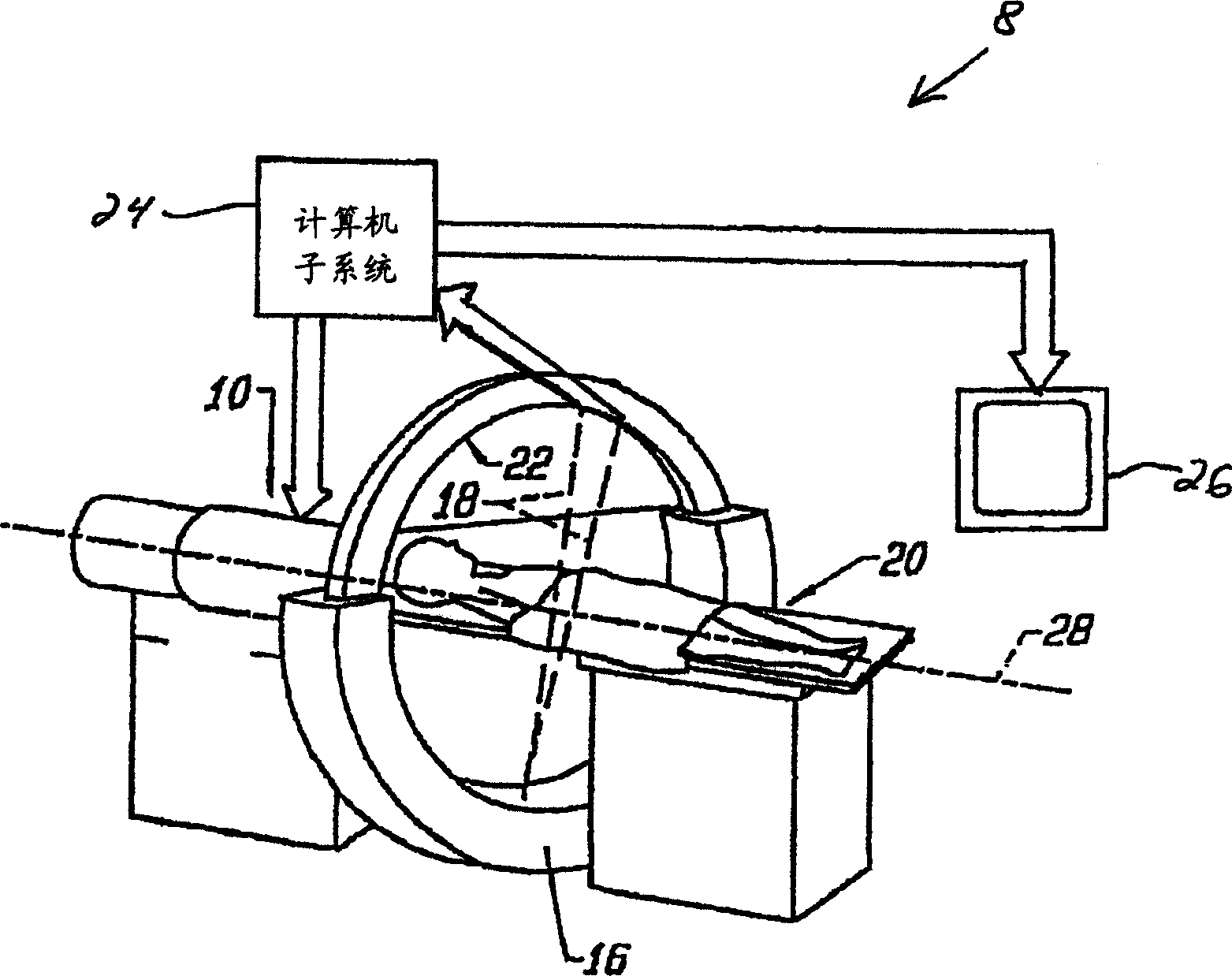
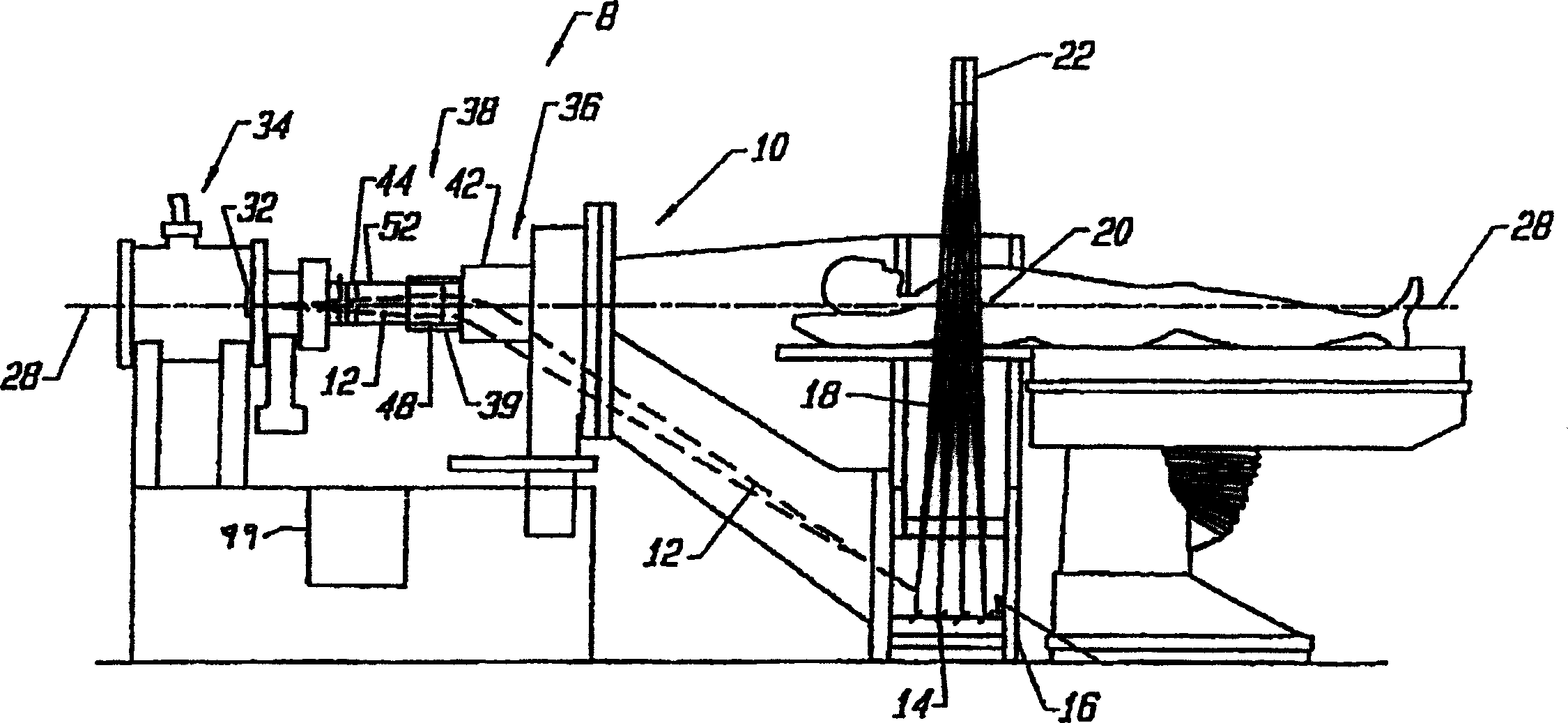
[0021] By way of example only, reference is made to certain embodiments of electron beam tomography (EBT) imaging systems in the following detailed description. It should be understood that imaging systems other than EBT imaging systems may be used with the present invention.

[0022] Before describing certain embodiments of the invention, it is helpful to understand the operation of an EBT imaging system. figure 1 with figure 2 An imaging system 8 formed in accordance with an embodiment of the present invention is illustrated. Such as figure 2 As shown, the system 8 includes a vacuum chamber 10 within which an electron beam 12 is generated at the cathode of an electron source 32 located in an upstream region 34 in response to a voltage (eg, -140 kV). The electron beam 12 is then controlled by an optical system 38 comprising a magnetic lens 39 and a deflection coil 42 to scan at least one half-ring target 14 located in the lower front portion 16 of the chamber 10 .

[00...


PUM


Abstract

Certain embodiments include a method and system for measuring and imaging local lung function. The method includes triggering an image scan of at least a lung cross-section of a patient, scanning the lung cross-section during at least one of an inspiration and an expiration of air by the patient to obtain lung image data and measuring a lung function during at least one of an inspiration and an expiration of air by the patient to obtain lung function data. The method may also include performing a preview scan of the patient to identify the lung cross-section for imaging. Additionally, the lung image data and lung function data may be combined for use in diagnosis of the patient. The lung image data and lung function data may also be output. The method may further include processing the lung function data to generate a plot of lung attenuation versus time.

Description

technical field [0001] This application is primarily concerned with imaging using electron beam tomography (EBT) scanners. In particular, the invention relates to measuring regional lung function using an EBT scanner. Background technique [0002] Medical diagnostic imaging systems encompass a variety of imaging modalities, such as x-ray systems, computed tomography (CT) systems, ultrasound systems, electron beam tomography (EBT) systems, magnetic resonance (MR) systems, and the like. Medical diagnostic imaging systems generate images of a subject, such as a patient, by exposing the subject to an energy source, such as x-rays passing through the patient, for example. The resulting image can be used in many ways. For example, it is possible to detect internal defects of the object under test and, in addition, to determine changes in the internal structure or arrangement. It can also represent the flow of liquid in the measured object. Furthermore, the images can show the ...

Claims


Application Information

Patent Timeline
IPC IPC(8): A61B5/091A61B6/00A61B6/03
CPCA61B6/541A61B6/032A61B6/4028
Inventor 戴维·G·希尔
Owner GE MEDICAL SYST GLOBAL TECH CO LLC
Why Eureka
Try Eureka