Method for separating and purifying stem cell of skin epidemis

A technology for separation and purification of epidermal stem cells, applied in the field of separation and purification of human and mammalian adult stem cells

Inactive Publication Date: 2005-01-12
NORTHWEST A & F UNIV
View PDF0 Cites 9 Cited by
  • Summary
  • Abstract
  • Description
  • Claims
  • Application Information

AI Technical Summary

Problems solved by technology

[0003] The purpose of the present invention is to overcome the disadvantage that the current enzymatic digestion method obtains mixed cell populations, and according to the characteristics of clonal growth of epidermal stem

Method used


Image

  • Method for separating and purifying stem cell of skin epidemis
  • Method for separating and purifying stem cell of skin epidemis
  • Method for separating and purifying stem cell of skin epidemis

Examples


Embodiment 1

[0020] Example 1: Separation, purification and expansion of epidermal stem cells from Guanzhong dairy goats

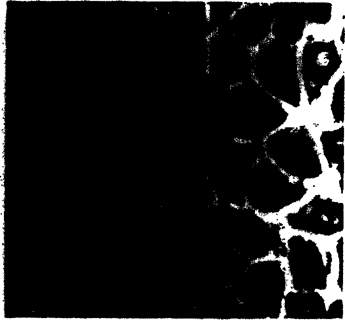
[0021] See attached picture, figure 1 Obtain the stem cell clone of the original epidermis of Guanzhong dairy goat by adopting the tissue block culture in the method of the present invention, X200; figure 2 It is the primary Guanzhong dairy goat epidermal stem cell integrin-α 6 Positive, X100; image 3 It is the first generation of Guanzhong Dairy Goat Epidermal Stem Cell β 1 -integrin positive, X200; Figure 4 It is the second generation of Guanzhong dairy goat epidermal stem cells positive for CK-19, X100; Figure 5 It is the second generation of Guanzhong dairy goat epidermal stem cells that are p63-positive X400.

[0022] In Guanzhong dairy goats (2.5-3.5-year-old ewes) where there are relatively few blood vessels in the middle of the ear, the hair is shaved and disinfected, and a piece of about 0.5×0.5cm in size is cut with ear size pliers. 2 Put the skin i...

Embodiment 2

[0025] Example 2: Separation, purification and expansion of human epidermal stem cells

[0026] Take a discarded piece after circumcision in the hospital, about 0.5×0.5cm in size 2 Put the skin in the normal saline containing penicillin (1000μ / ml) and bring it back to the sterile room, and wash it several times with PBS (1) containing penicillin to remove blood stains. Separate the skin from the subcutaneous tissue under a dissecting microscope, and cut the skin into 0.1-0.2×0.1-0.2cm 2 Small pieces of different sizes are placed on glass plates (φ=5.0cm) with the skin side up, 3-4 pieces per plate, and an appropriate amount of medium (Medium199+15-20% NBS+5-8μg / ml insulin+ 0.5μg / ml hydrocortisone + penicillin 100u / ml), placed in CO 2 Incubator (5% CO 2 , saturated humidity, 37°C), and the medium was changed every two days. After 3-4 days, epithelioid cells grow like paving stones from the edge of the tissue block. After 7-12 days, there are small round cells aggregated on ...


PUM


Abstract

This invention discloses a method for separating and purifying skin caticular dry cells which adopts a self designed culture medium to suppress growth of fibrablasts and unform epithelioid cells grow around the tissue block to induce the cells to move out of the original micro-environment and gather again and grow on the new grown epithelioid cell layer. These clone grown cells are obviously different in morphology from its dependent trophocytes and can be selected with the help of dissection mirror and glass pins and nurtured for expansion independently. The acquired cells are tested by the generally acknowledged skin dry cells molecular label: ck19, integrin-beta1, P63, integrin-alpha 6 are positive which means the selected ones are purified skin dry cells.

Description

technical field [0001] The invention belongs to the technical field of separation and purification of human and mammalian adult stem cells, and specifically relates to a method for separation, purification and expansion of human and mammalian skin epidermal stem cells. Background technique [0002] Epidermal stem cells s ,ESC s ) is a stem cell with self-proliferation and multi-directional differentiation potential. Its normal proliferation and differentiation are the basic requirements for maintaining the structural and functional integrity of the skin and its appendages (sweat glands, hair, sebaceous glands). Under physiological conditions, epidermal stem cells differentiate into a stem cell and a transient expanding cell through asymmetric division. s TA cells), and TA cells differentiate into post-mitotic cells after multiple divisions s ) and terminally-differentiated cells s ). To supplement the continuous renewal needs of epidermal cells. Studies have shown tha...

Claims


Application Information

Patent Timeline
IPC IPC(8): C12N5/071C12N5/074C12Q1/24
Inventor 杨学义窦忠英
Owner NORTHWEST A & F UNIV
Why Eureka
Try Eureka