Method for inducing fibroblast to form cartilage cells

A technology for fibroblasts and chondrocytes, which is applied in the fields of biology and tissue engineering, and can solve problems such as making chondrocytes

Active Publication Date: 2005-08-31
上海组织工程研究与开发中心
View PDF0 Cites 4 Cited by
  • Summary
  • Abstract
  • Description
  • Claims
  • Application Information

AI Technical Summary

Problems solved by technology

However, so far, there is no report on the production o

Method used


Image

  • Method for inducing fibroblast to form cartilage cells
  • Method for inducing fibroblast to form cartilage cells
  • Method for inducing fibroblast to form cartilage cells

Examples


Embodiment 1

[0085] Fibroblast culture and passaging

[0086]Under sterile conditions, the skin and dermis tissue excised during circumcision was taken, cut into pieces and transferred to a centrifuge tube, and digested by adding 0.1% type I collagenase (WORTHINGTON company) about 10 times the volume of the tissue, at 37°C for 2 hours, After filtering with a 100-mesh cell strainer, centrifuge at 1000 rpm for 5 minutes, discard the supernatant, add 10% FBS DMEM culture solution (GIBCO) to mix the cells, and dilute the cells at 2×10 4 / cm 2 Inoculated in 100mm culture dish (FALCON, FranklinLakes NJ) for culture, when the cells were confluent to 80%-90%, subcultured, the subculture density was 1×10 4 / cm 2 . After in vitro expansion to the second generation, the experimental grouping was carried out.

Embodiment 2

[0088] Induction of Chondrocytes in Monolayer Culture

[0089] In this example, chondrocytes were induced by monolayer culture cell induction method. Methods as below:

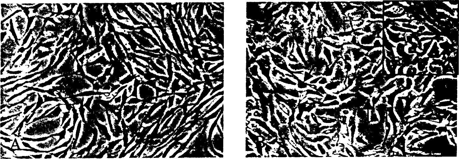
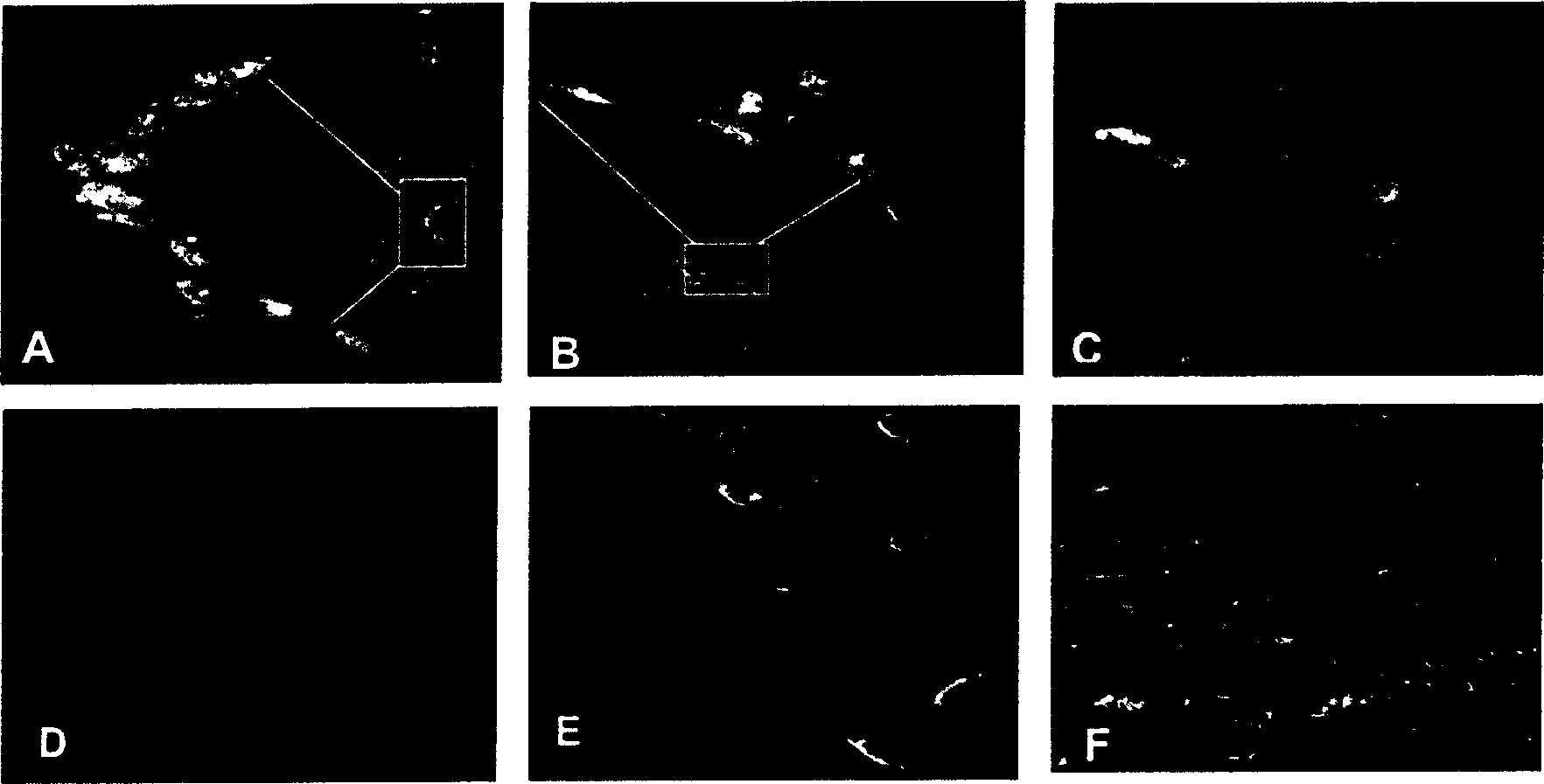
[0090] Dilute CDMP1 growth factor (Research Diagnostics) to 100 μg / ml with 0.1% BSA, add F-12 (Gibco Company) culture solution containing 10% FBS to make the final concentration 100 ng / ml, and prepare cell induction solution. The second-generation fibroblasts were inoculated for 6-12 hours, and the cells were induced. The culture medium was changed every three days, and the induction was carried out for 7 days (at 37°C, 5% CO 2 , saturated humidity), and isolated to obtain chondrocytes.

Embodiment 3

[0092] Centrifuge Tube Culture Cell Induction

[0093] In this example, chondrocytes were induced by monolayer culture cell induction method. Methods as below:

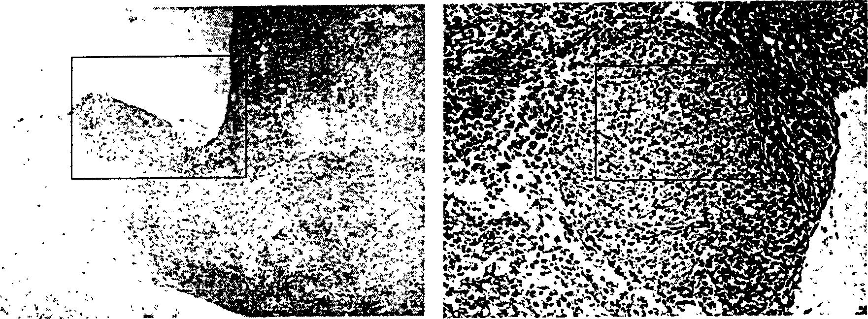
[0094] The preparation of the induction solution was the same as in Example 2. Collect the fibroblasts that have been passaged to the 6th passage and centrifuge at 1500 rpm for 5 minutes, discard the supernatant, add cell induction solution, shake the cell mass, induce for 7 days, and obtain the chondrocyte mass.

[0095] Paraffin-embedded and sectioned for identification by Masson's Trichrome, Alcian Blue and immunohistochemical staining.


PUM


Abstract

A process for inducing fibroblast to become cartilage cell includes culturing the fibroblasts in the culture liquid contaniing the cartilage form generating protein for 3-30 days to generate cartilage cells, and separating them for culture liquid.

Description

technical field [0001] The present invention relates to the fields of biology and tissue engineering, and more particularly to a method for inducing fibroblasts to form chondrocytes. Background technique [0002] As a kind of connective tissue, cartilage mainly supports, protects, and disperses stress. Histologically, it can be divided into three types: hyaline cartilage (articular cartilage, costal cartilage, nasal cartilage, tracheal cartilage), elastic cartilage (epiglottic cartilage, ear cartilage), and fibrocartilage (meniscus). Histologically, they are all single tissues containing only chondrocytes, without blood vessels inside, and their nutrition mainly depends on the penetration of interstitial fluid. [0003] Structurally, chondrocytes are wrapped in the cartilage lacuna, and the thick cartilage capsule around it acts as a natural barrier to separate chondrocytes from surrounding tissues. Therefore, cartilage tissue has weak antigenicity, is not easy to be recog...

Claims


Application Information

Patent Timeline
IPC IPC(8): C12N5/06C12N5/08
Inventor 崔磊曹谊林刘伟尹烁
Owner 上海组织工程研究与开发中心
Why Eureka
Try Eureka