Confocal endoscope mini-microscope objective lens probe

A microscopic objective lens and endoscope technology, which is applied in the field of devices in the field of optical technology, can solve the problems of small working distance, large volume, and small field of view of the microscopic objective lens, and achieve noise suppression, large numerical aperture, and small overall size Effect

Inactive Publication Date: 2006-03-08
SHANGHAI JIAO TONG UNIV
View PDF1 Cites 24 Cited by
  • Summary
  • Abstract
  • Description
  • Claims
  • Application Information

AI Technical Summary

Problems solved by technology

"Its shortcomings are: 1. This design is used for general microscopes, which are relatively large in size and cannot enter the human body through the endoscope system, nor can it be coupled with imaging fiber bundles; 2. The working distance of this microscope objective lens It is also very small, generally around 100-200 microns; 3. The field of view of this microscope objective is also relatively small

Method used


Image

  • Confocal endoscope mini-microscope objective lens probe
  • Confocal endoscope mini-microscope objective lens probe

Examples


Embodiment Construction

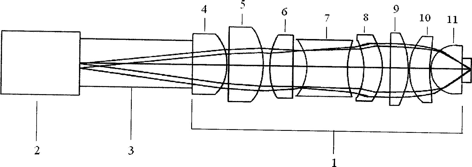
[0016] like figure 1 As shown, the present invention includes: a microscopic objective lens group 1, an imaging fiber bundle 2 and a refractive index matching liquid 3, and the microscopic microscopic objective lens group 1 includes: a first lens element 4, a second lens element 5, a third Lens element 6 , fourth lens element 7 , fifth lens element 8 , sixth lens element 9 , seventh lens element 10 and eighth lens element 11 , the first lens element 4 is arranged at the front of the microscopic objective lens group 1 5.63mm behind the imaging fiber bundle 2, the second lens element 5 is located 0.097mm after the first lens element 4, the third lens element 6 is located 0.257mm after the second lens element 5, and the fourth lens element 7 is located 0.52mm before , the fifth lens element 8 is placed 0.597mm after the fourth lens element 7, the sixth lens element 5 is placed 0.437mm after the fifth lens element 8, 0.05mm before the seventh lens element 10, and the eighth lens e...


PUM


Abstract

A micro object lens probe for confocal endoscopes, which contains micro object lens group, imaging optical beam and refractive index matching solution filled between imaging optical fiber beam and micro object lens group, said imaging optical fiber beam set before micro object lens group whose pupil located infinite farness, said micro object lens group has 1.0 hole diameter matched with that of single optical fiber of imaging optical fiber beam.

Description

technical field [0001] The invention relates to a device in the field of optical technology, in particular to a confocal endoscope microscopic objective lens probe. Background technique [0002] At present, the confocal system has high imaging contrast and resolution due to its clear imaging, continuous slice scanning and image reorganization, multi-label technology, in vivo observation and obtaining quantitative information, etc., and is widely used in various fields of medicine. big effect. However, when observing tissue, the system must take out the internal organ tissue of the human body, and then perform imaging and analysis, which not only increases the pain of the patient, but also prolongs the diagnosis cycle, so it cannot be popularized clinically. This limitation prompted the development of fiber-optic confocal microscopy. The optical fiber creates a flexible form of connection between the objective of the confocal microscope and the rest of the system, which all...

Claims


Application Information

Patent Timeline
Patent Type & Authority Applications(China)
IPC IPC(8): G02B23/26G02B9/64G02B13/26G02B21/02
Inventor 王成任秋实余文娟
Owner SHANGHAI JIAO TONG UNIV
Why Eureka
Try Eureka