Method for submit inspection result in three-dimensional imaging especially in mammary glad X-ray photograph

A technology of inspection results and three-dimensional imaging, which is used in mammography, instruments for radiological diagnosis, image enhancement, etc. The effect of saving work

Inactive Publication Date: 2006-04-26
SIEMENS AG
View PDF1 Cites 4 Cited by
  • Summary
  • Abstract
  • Description
  • Claims
  • Application Information

AI Technical Summary

Problems solved by technology

Therefore, this imaging examination method is mainly used in screening, that is, in the continuous examination of patients with a patient volume of more than 50 patients per day, which is no longer easily competent for doctors in terms of the cost of examination and diagnosis.

Method used


Image

  • Method for submit inspection result in three-dimensional imaging especially in mammary glad X-ray photograph
  • Method for submit inspection result in three-dimensional imaging especially in mammary glad X-ray photograph
  • Method for submit inspection result in three-dimensional imaging especially in mammary glad X-ray photograph

Examples


Embodiment Construction

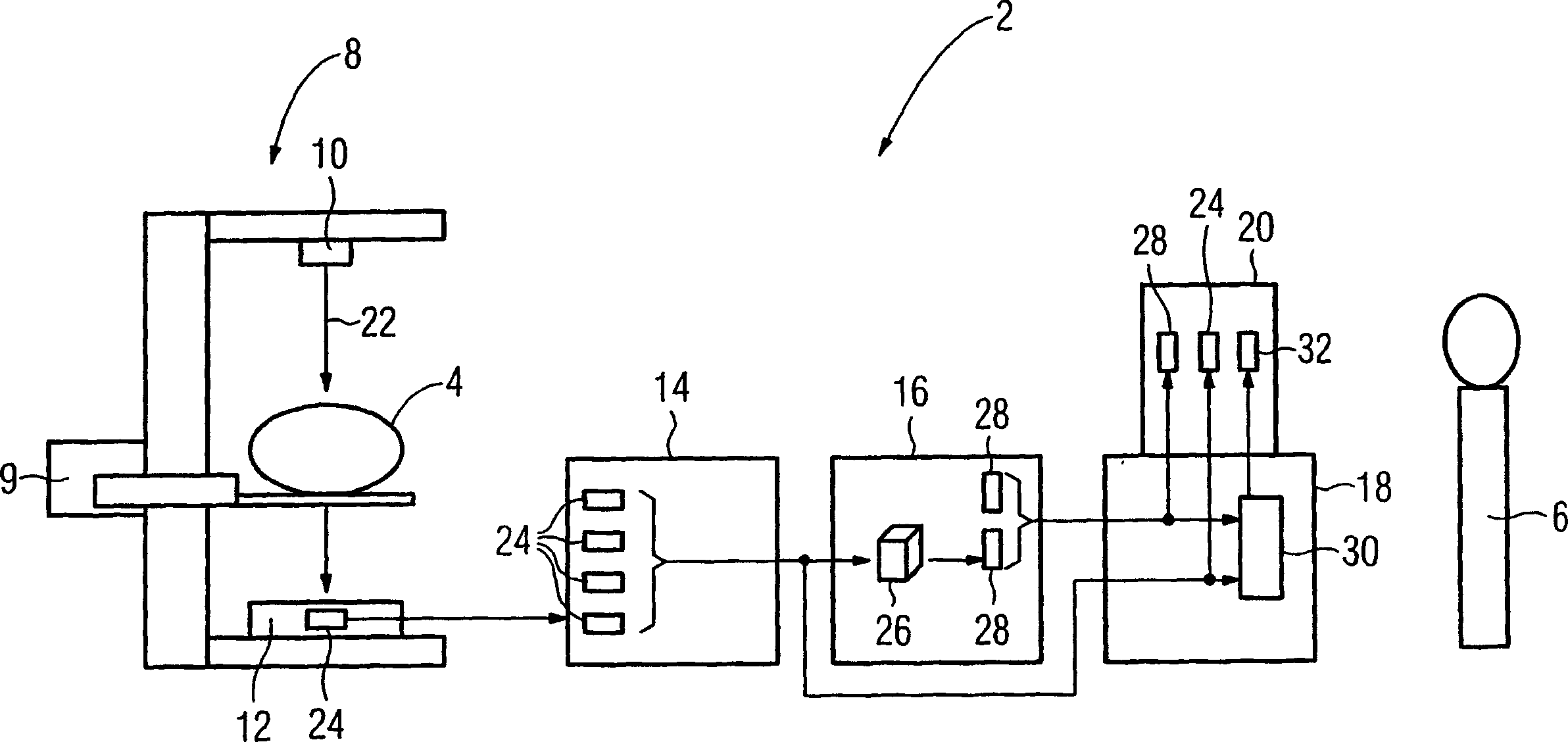
[0031] figure 1 A tomograph 2 is shown that produces examination results in three-dimensional imaging or within the framework of tomography, including a patient 4 to be examined and a doctor 6 performing the examination. The tomograph 2 comprises an x-ray device 8, a tomograph computer 16 and an examination and diagnosis computer 18 with a display 20, wherein the x-ray device 8 has an x-ray source 10, a mechanical translation device 9 and a digital detector 12 and a computer 14 assigned to the X-ray system 8 .

[0032] At a so-called acquisition workstation in the form of a computer 14 which is used as a workstation by the medical staff or doctor 6 , the doctor initiates an examination of the patient, for example a mammography of a woman's breast. Next, the patient 4 is fluoroscopyed with the x-rays 22 emitted from the x-ray source 10, and in the process a single projection or projection image 24 is produced in the detector 12, wherein the patient 4 and / or The relative posit...


PUM


Abstract

The invention relates to a method for producing examination results in three-dimensional imaging, in particular mammograms, in which a projection image of an object to be examined is produced and stored electronically. Tomographic images are reconstructed from projected photographs using reconstruction methods. The doctor observes the tomographic image and gives inspection results, and marks positive inspection results in the tomographic image with a first marker. The CAD system gives inspection results to the tomographic image, and marks positive inspection results in the tomographic image with a second marker. A tomographic image different from the first marker and the second marker will be re-observed by a doctor and an examination result will be given.

Description

technical field [0001] The invention relates to a method for producing examination results in three-dimensional imaging, in particular in mammograms. Background technique [0002] Imaging methods are of increasing importance in medical technology. The inspection object is a human body or an animal body. Classical methods such as X-ray techniques have long existed, in which a two-dimensional image of the body region to be examined is produced. Furthermore, there are modern methods such as nuclear spin tomography or computed tomography, which also produce three-dimensional images or, by tomography, approximately three-dimensional images. [0003] In the digital tomosynthesis method known, for example, from DE 19842944, a digital projection x-ray system is used to obtain three-dimensional information which is provided in the form of reconstructed slices. [0004] In practice, classical tomography (so-called “tomography”) has proven to be the forerunner of digital tomography ...

Claims


Application Information

Patent Timeline
Patent Type & Authority Applications(China)
IPC IPC(8): A61B6/00A61B6/03G06T17/00
CPCA61B6/502G06T7/0012G06T2207/30068
Inventor 托马斯·默特尔迈耶
Owner SIEMENS AG
Why Eureka
Try Eureka