

Detection method of hepatitis B virus genome drug resistance mutation
A technology of hepatitis B virus and mutant type, which is applied in biochemical equipment and methods, and microbial measurement/inspection, etc., can solve the problems of cumbersome operation, high cost, and unfavorable development of primary hospitals, so as to improve the diffusion rate and simple operation Effect
- Summary
- Abstract
- Description
- Claims
- Application Information
AI Technical Summary
Problems solved by technology
Method used
Image

Examples
Embodiment 1
[0090] Embodiment 1: the extraction of serum DNA
[0091] Aseptically collect about 1ml of venous blood from the hepatitis B patient to be tested, let it stand at room temperature and 4°C for 2 hours and 1 hour respectively, then centrifuge (8,000rpm, 5 minutes) to separate and collect 200μl of serum sample. Then take 40 μl of serum sample, add an equal amount of DNA extraction solution to it, mix thoroughly and place in a boiling water bath for 10 minutes. In order to ensure that the virus particles contained in the serum can be fully lysed, the mixture was further left to stand at 4° C. for 10 hours. It was then centrifuged (10,000 rpm, 5 minutes) and 2 μl of the supernatant was collected and used for further PCR reactions.
Embodiment 2
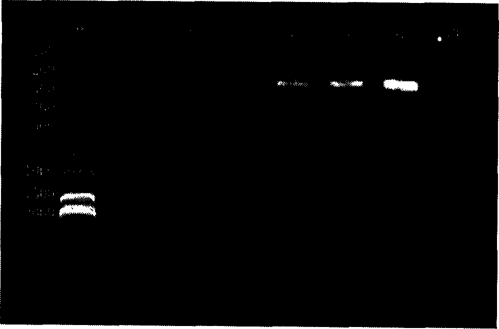
[0092] Embodiment 2: PCR amplification of the fragment comprising YMDD motif
[0093] Take several tubes of single-person PCR reaction solution, add 1 μl UDG enzyme to each tube, and then directly add 2 μl template (or negative and positive standards), mix well and then centrifuge briefly (3 seconds). Then put each reaction tube into the PCR instrument, pretreat at 50°C for 3 minutes, and amplify according to the following conditions: 94°C for 5 minutes, then press 93°C for 30 seconds, 55°C for 30 seconds, and 72°C for 40 seconds, a total of 35 cycles, A final 72°C extension was performed for 7 minutes. The amplified products were detected by 2% agarose gel electrophoresis (see attached figure 1 ). The PCR amplification system used included: 1×qualitative PCR buffer, 0.2 mM dNTPs, 2 U Taq enzyme, 0.2 μM primers 1 (SEQ ID NO: 1) and 2 (SEQ ID NO: 2).
Embodiment 3
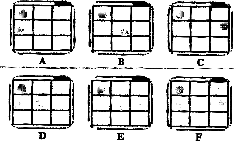
[0094] Example 3: Reverse dot blot detection of samples of three known genotypes
[0095]Before hybridization, preheat hybridization solution I (2×SSC-0.1% SDS) and hybridization solution II to 44°C for use. Take three 1.5ml centrifuge tubes according to the number of samples to be tested, add 0.5ml hybridization solution I to each tube and preheat to 44°C. After the PCR amplification product was denatured at 95° C. for 10 minutes, it was placed in an ice-water mixture for 4 minutes. Then take 1000 μl hybridization solution I+2 μl solution I (1000:2) mixed solution as the binding solution and store it at 4°C for later use, and take the mixture of solution II: solution III: solution IV (1900:200:1) (1.9ml solution II+ 0.2ml of solution III + 1 μl of solution IV) is used as a chromogenic solution and stored away from light for later use.
[0096] Before hybridization, fill the reaction chamber with distilled water, place the metal perforated plate, and turn on the negative pre...
PUM
 Abstract
Description
Claims
Application Information