Method and product for localized or spatial detection of nucleic acid in a tissue sample
a nucleic acid and tissue sample technology, applied in the field of localized or spatial detection of nucleic acid in tissue samples, can solve the problems of inability to achieve global transcriptome analysis with spatial resolution, low precision, and high throughput methods for studying transcriptional activity with high resolution in intact tissues, etc., to achieve the effect of multiple analyses
- Summary
- Abstract
- Description
- Claims
- Application Information
AI Technical Summary
Benefits of technology
Problems solved by technology
Method used
Image
Examples
example 1
[0361]Preparation of the Array
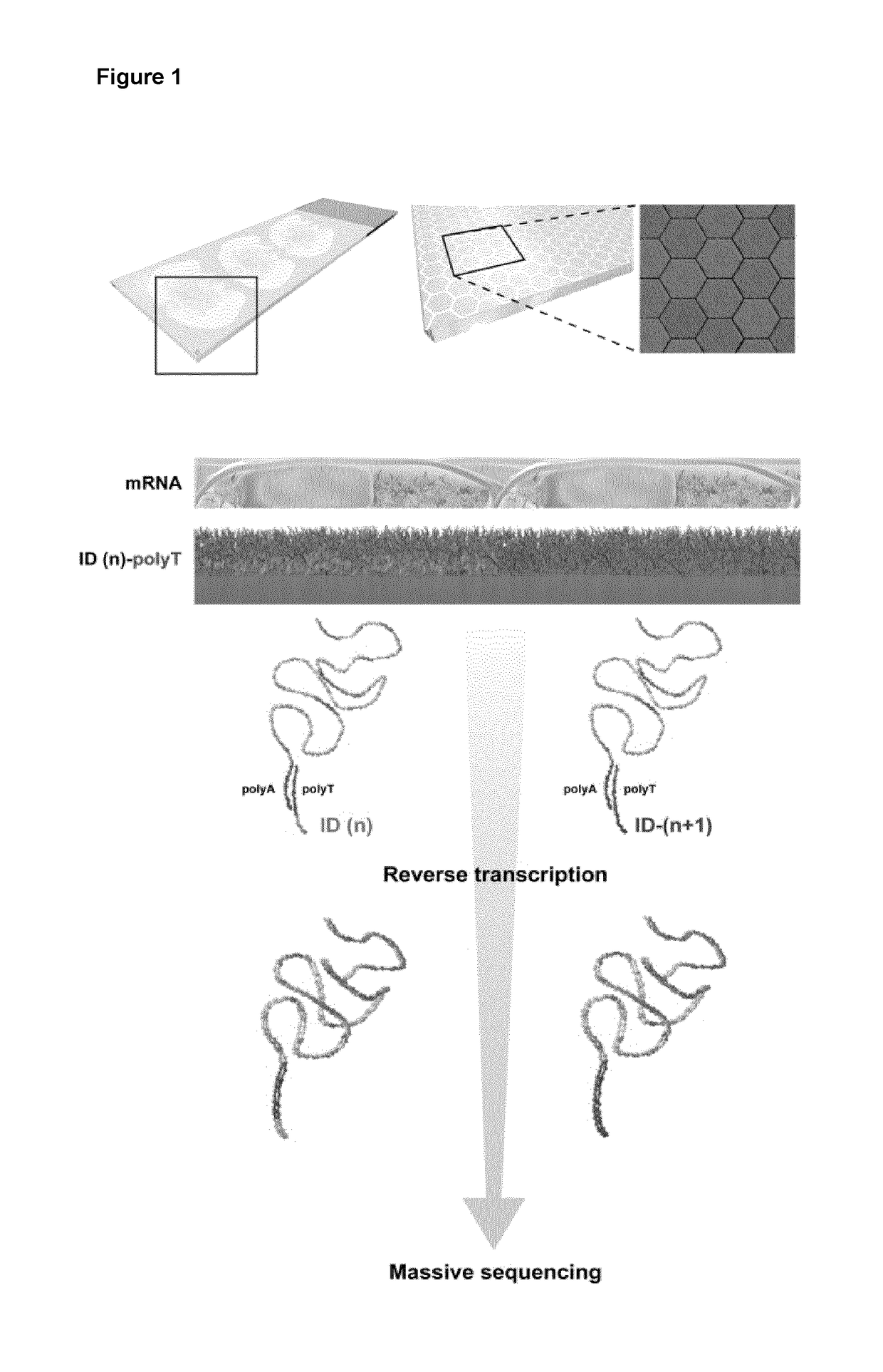
[0362]The following experiments demonstrate how oligonucleotide probes may be attached to an array substrate by either the 5′ or 3′ end to yield an array with capture probes capable of hybridizing to mRNA.
[0363]Preparation of in-house Printed Microarray with 5′ to 3′ Oriented Probes
[0364]20 RNA-capture oligonucleotides with individual tag sequences (Tag 1-20, Table 1 were spotted on glass slides to function as capture probes. The probes were synthesized with a 5′-terminus amino linker with a C6 spacer. All probes where synthesized by Sigma-Aldrich (St. Louis, Mo., USA). The RNA-capture probes were suspended at a concentration of 20 μM in 150 mM sodium phosphate, pH 8.5 and were spotted using a Nanoplotter NP2.1 / E (Gesim, Grosserkmannsdorf, Germany) onto CodeLink™ Activated microarray slides (7.5 cm×2.5 cm; Surmodics, Eden Prairie, Minn., USA). After printing, surface blocking was performed according to the manufacturer's instructions. The probes were pr...
example 2
[0433]FIGS. 8 to 12 show successful visualisation of stained FFPE mouse brain tissue (olfactory bulb) sections on top of a bar-coded transcriptome capture array, according to the general procedure described in Example 1. As compared with the experiment with fresh frozen tissue in Example 1, FIG. 8 shows better morphology with the FFPE tissue. FIGS. 9 and 10 show how tissue may be positioned on different types of probe density arrays.
example 3
[0434]Whole Transcriptome Amplification by Random Primer Second Strand Synthesis Followed by Universal Handle Amplification (Capture Probe Sequences Including Tag Sequences Retained at the End of the Resulting dsDNA)
[0435]Following capture probe release with uracil cleaving USER enzyme mixture in PCR buffer (covalently attached probes)
OR
[0436]Following capture probe release with heated PCR buffer (hybridized in situ synthesized capture probes)
[0437]1 μl RNase H (5 U / l) was added to each of two tubes, final concentration of 0.12 U / μl, containing 40 μl 1× Faststart HiFi PCR Buffer (pH 8.3) with 1.8 mM MgCl2 (Roche, www.roche-applied-science.com), 0.2 mM of each dNTP (Fermentas, www.fermentas.com), 0.1 μl / μl BSA (New England Biolabs, www.neb.com), 0.1 U / μl USER Enzyme (New England Biolabs), released cDNA (extended from surface probes) and released surface probes. The tubes were incubated at 37° C. for 30 min followed by 70° C. for 20 min in a thermo cycler (Applied Biosystems, www.appl...
PUM
| Property | Measurement | Unit |
|---|---|---|
| area | aaaaa | aaaaa |
| sizes | aaaaa | aaaaa |
| feature sizes | aaaaa | aaaaa |
Abstract
Description
Claims
Application Information
 - R&D
- Intellectual Property
- Life Sciences
- Materials
- Tech Scout
- Unparalleled Data Quality
- Higher Quality Content
- 60% Fewer Hallucinations
Browse by: Latest US Patents, China's latest patents, Technical Efficacy Thesaurus, Application Domain, Technology Topic, Popular Technical Reports.
© 2025 PatSnap. All rights reserved.Legal|Privacy policy|Modern Slavery Act Transparency Statement|Sitemap|About US| Contact US: help@patsnap.com


