2D/3D registration

a technology of 2d/3d and registration, applied in the field of 2d/3d registration, can solve the problems of prejudiced motion estimation and unnecessary computational operations, and achieve the effects of avoiding complex and time-consuming volume analysis, significant improvement of real-time capability of registration, and fundamental increase in computational efficiency

Active Publication Date: 2018-08-28
SIEMENS HEALTHCARE GMBH
View PDF4 Cites 0 Cited by
  • Summary
  • Abstract
  • Description
  • Claims
  • Application Information

AI Technical Summary

Benefits of technology

[0022]Since operations are image-based and performed in the 2D domain, the approach according to one or more of the present embodiments is particularly suitable for realizing a multiple resolution scheme in which progress is made successively toward lower resolutions. Overall, the method according to one or more of the present embodiments offers a more efficient possibility for finding contour points (and the associated three-dimensional starting points in the three-dimensional image dataset), which may be utilized, for example, in connection with the known point-to-plane correspondence model described by the cited publications. The proposed operation on the gradient comparison images avoids a complex and time-consuming volume analysis. If, as already mentioned, a multiple resolution scheme is used or if the process is essentially initiated based on a coarser resolution, the selected contour points are sparsely distributed; overlaid contour points and redundant calculations are avoided. Accordingly, computational efficiency is fundamentally increased, and consequently, the real-time capability of the registration is significantly improved.
[0027]One embodiment provides that the gradient comparison image is determined by applying a render algorithm to the gradient dataset. In this case, one result channel of the render algorithm contains a normalized angular value as a direction indication of the gradient in the image plane and a further result channel contains a measure for the magnitude of the gradient in the image plane. In the present method also, therefore, the gradient in the image plane is not determined from digitally reconstructed radiograph (DRR) images forward-projected from the three-dimensional image dataset, but is rendered directly. This prevents a “false” gradient in the image plane by direct manipulation of the three-dimensional gradient of the gradient dataset, whereas “false” gradients may occur if two-dimensional image gradients are determined from DRR images. An example of this is the occurrence of false edges, which are produced due to artificially resulting limitations during the partitioning into depth intervals. A further advantage of direct volume rendering from the gradient dataset is that the render operation may be carried out in a single step on a graphical processing unit (GPU). In one embodiment, the render algorithm is executed by a GPU. An extremely fast and efficient calculation is possible by this, since hardware that is specifically configured for performing such render operations is used.
[0032]In one embodiment of the method, the gradient x-ray image and the gradient comparison image are initially determined in a coarser resolution with larger image elements than the x-ray image. The determination of the motion parameters and the correction of the test transformation are repeated successively in order at least to provide a better resolution (e.g., up to the resolution of the x-ray image). As has already been indicated, the image-based analysis conducted in order to find contour points is particularly suited to such a multiple resolution scheme, by which consideration is initially given to larger image elements that combine smaller image elements of the size of the image elements of the x-ray image. This has several advantages. In this way, the size of the area in which movements relative to the three-dimensional image dataset are captured may be increased, since the algorithm initially reacts less sensitively. The robustness of the procedure is therefore increased significantly. A type of “pyramid representation” is therefore given, where, for example, resolutions may be increased from 64×64 image elements, through 128×128 and 256×256 image elements, to 512×512 image elements. In this way, the accuracy of the determination of the motion parameters, and consequently the registration transformation, is improved incrementally.
[0035]The method according to one or more of the present embodiments reduces the computational overhead by mapping the reference structures as a sparse representation and enabling operation to be performed on a two-dimensional basis. With the iterative approach, work is focused, for example, on the starting points and the contour points associated therewith. The comparison method is henceforth performed locally. Once a 2D / 3D correspondence has been found locally, the 2D / 3D correspondence may not be lost in further iterations. In the proposed approach, a gradient comparison image is generated directly from the three-dimensional image dataset. The search area for the 2D / 3D tracking may be based on the image contents (e.g., as explained in more detail below, on the gradient direction). In this way, the method provides valid registrations both at the start of a medical intervention and during the further course thereof, as well as in pauses during the acquisition of the x-ray images. Since the three-dimensional image dataset is generally taken as the basis, there is also no further accumulation of tracking errors, because an accurate registration between the three-dimensional image dataset and the two-dimensional x-ray image is established immediately. In a certain manner, the method therefore provides a natural initialization (e.g., reinitialization) for the differential motion compensation (e.g., when the initial transformation is based on the registration transformation established in the previous time increment). In this way, the motion compensation framework becomes more reliable, since no errors accumulate and a refinement of the registration ultimately occurs automatically when it becomes necessary. Persons performing a medical intervention are able to concentrate more on the intervention procedure without being interrupted by misregistrations and the like.
[0037]A suitable initial transformation may already be determined with sufficient accuracy from a known position of the patient relative to the x-ray device acquiring the x-ray image. When positioning patients inside an x-ray device having, for example, a C-arm (e.g., for a medical intervention), the position of the patient, and therefore the location of the target region, is in most cases already known very precisely, while the acquisition geometry and the geometric properties of the x-ray device are also available. Thus, given a known acquisition geometry, the initial transformation may be estimated with sufficient accuracy so as to lead to sufficiently reliable success in the 2D / 3D registration (e.g., given suitable parameterization of the registration algorithm provided (e.g., insofar as the extent of the environments and / or the search area are concerned).
[0039]As has already been mentioned, the present method is suitable, for example, for the fluoroscopic x-ray monitoring of, for example, minimally invasive medical interventions. In this context, it is beneficial if a time series of x-ray images is acquired (e.g., for the purpose of monitoring a medical intervention). A registration is performed for each x-ray image, and the registration transformation determined for the x-ray image acquired immediately beforehand is used in each case as the initial transformation. Since there is typically only a short time interval between the acquisitions of x-ray images succeeding one another in time, particularly strong movements are unlikely to occur in the target region, so that the final registration transformation in relation to the previously acquired x-ray image is suitable as an initial transformation for the next registration process. A quality of the initial transformation known in this way may also be used, as already mentioned, in order to adapt parameters, for example, with regard to the described 2D / 3D tracking, such that an embodiment provides that in the case of a registration transformation determined for an x-ray image acquired immediately beforehand as the initial transformation, a smaller environment and / or a smaller number of test points are / is chosen than in the case of some other initial transformation. If, for example, the x-ray image has a resolution of 512*512 pixels, an environment equal in size to 20*20 pixels or image elements may be applied in the case of a large assumed deviation of the initial transformation (e.g., at the time of the first determination of a registration transformation from a coarse transformation; initially, while the environment used for the comparison may be reduced to 5*5 pixels or image elements when registration transformations of preceding x-ray images are used). Similarly, a smaller number of test points or a smaller search area may be chosen.

Problems solved by technology

In the dynamic registration approach described therein, the selection of the starting points (and consequently contour points) was namely non-trivial for a variety of reasons (e.g., because the projected contour points typically cover the regions of two-dimensional interest in a redundant manner, with the result that unnecessary computational operations are required, and because a selection of the projected contour points performed anyway does not ensure a useful distribution of the contour points, with the result that a prejudiced motion estimation may occur due to loss of information).

Method used


Image

  • 2D/3D registration
  • 2D/3D registration
  • 2D/3D registration

Examples


Embodiment Construction

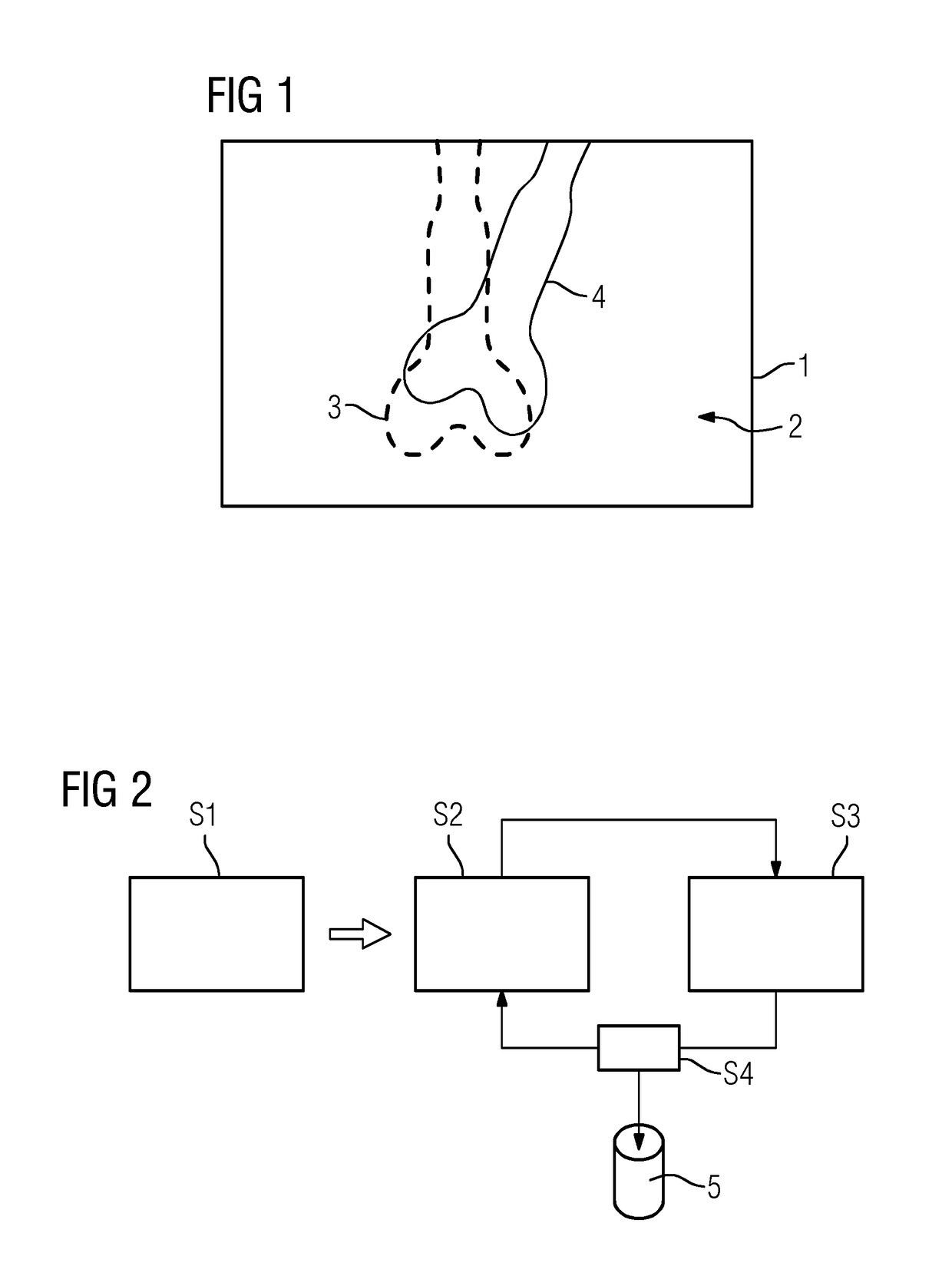
[0077]FIG. 1 shows, by way of example and in a schematic representation, an overlay image 1 from a three-dimensional image dataset and an x-ray image. The x-ray image 2 forms the basis onto which the information 3 of the three-dimensional image dataset is superimposed as an overlay. A shadow-like, low-resolution anatomical structure 4 (e.g., bone) is discernible in the x-ray image 2. The information 3 relating to the same anatomical structure 4, the overlaying being brought about using a coarse initial transformation (e.g., hence, a coarse 2D / 3D registration), is superimposed from the three-dimensional image dataset. It is apparent that the anatomical structure according to the information 3 is slightly skewed and translated relative to the visible anatomical structure 4 of the x-ray image 2. The below-described exemplary embodiment of the method aims to determine a registration transformation that leads to an accurate overlaying of said anatomical structures, and hence, to an accur...


PUM


Abstract

A method includes, following specification of an initial transformation as a test transformation that is to be optimized, determining a 2D gradient x-ray image and a 3D gradient dataset of the image dataset, carrying out, for each image element of the gradient comparison image, a check for selection as a contour point, and determining an environment best corresponding to a local environment of the contour point and extending around a comparison point in the gradient x-ray image for all contour points in the at least one gradient comparison image. Local 2D displacement information is determined by comparing the contour points with the associated comparison points, and motion parameters of a 3D motion model describing a movement of the target region between the acquisition of the image dataset and the x-ray image are determined from the displacement information and a registration transformation describing the registration.

Description

[0001]This application claims the benefit of DE 10 2016 202 512.0, filed on Feb. 18, 2016, which is hereby incorporated by reference in its entirety.BACKGROUND[0002]The present embodiments relate to registering a three-dimensional image dataset of a target region of a patient with a two-dimensional x-ray image of the target region recorded in an acquisition geometry.[0003]In many problematic medical situations, whether it be in diagnostics or when performing minimally invasive interventions, for example, both three-dimensional image data in the form of an image dataset (e.g., a computed tomography or magnetic resonance dataset) and two-dimensional fluoroscopic images (e.g., typically x-ray images) are available for the target region of interest of the patient. In such situations, it is desirable to be able to carry out a joint evaluation of both the three-dimensional image dataset and the two-dimensional x-ray image of the target region (e.g., in the form of a fusion image or by int...

Claims


Application Information

Patent Timeline
Patent Type & Authority Patents(United States)
IPC IPC(8): G06K9/00A61B6/00G06T7/207G06T7/564G06T7/38G06T7/00G06T19/00G06T11/00G06T7/246G06T7/32G06T7/37G06T7/269
CPCG06T7/37A61B6/5264G06T7/0016G06T7/207G06T7/248G06T7/269G06T7/32G06T7/38G06T7/564G06T11/005G06T11/006G06T19/003A61B6/5211G06T2207/30008G06T2200/04G06T2200/28G06T2207/10028G06T2207/10081G06T2207/10088G06T2207/10121G06T2207/20016G06T2207/20221G06T2207/10116G06T7/33A61B6/032A61B6/487A61B6/5229A61B6/5235
Inventor SCHAFFERT, ROMANWANG, JIANBORSDORF, ANJA
Owner SIEMENS HEALTHCARE GMBH
Why Eureka
Try Eureka