

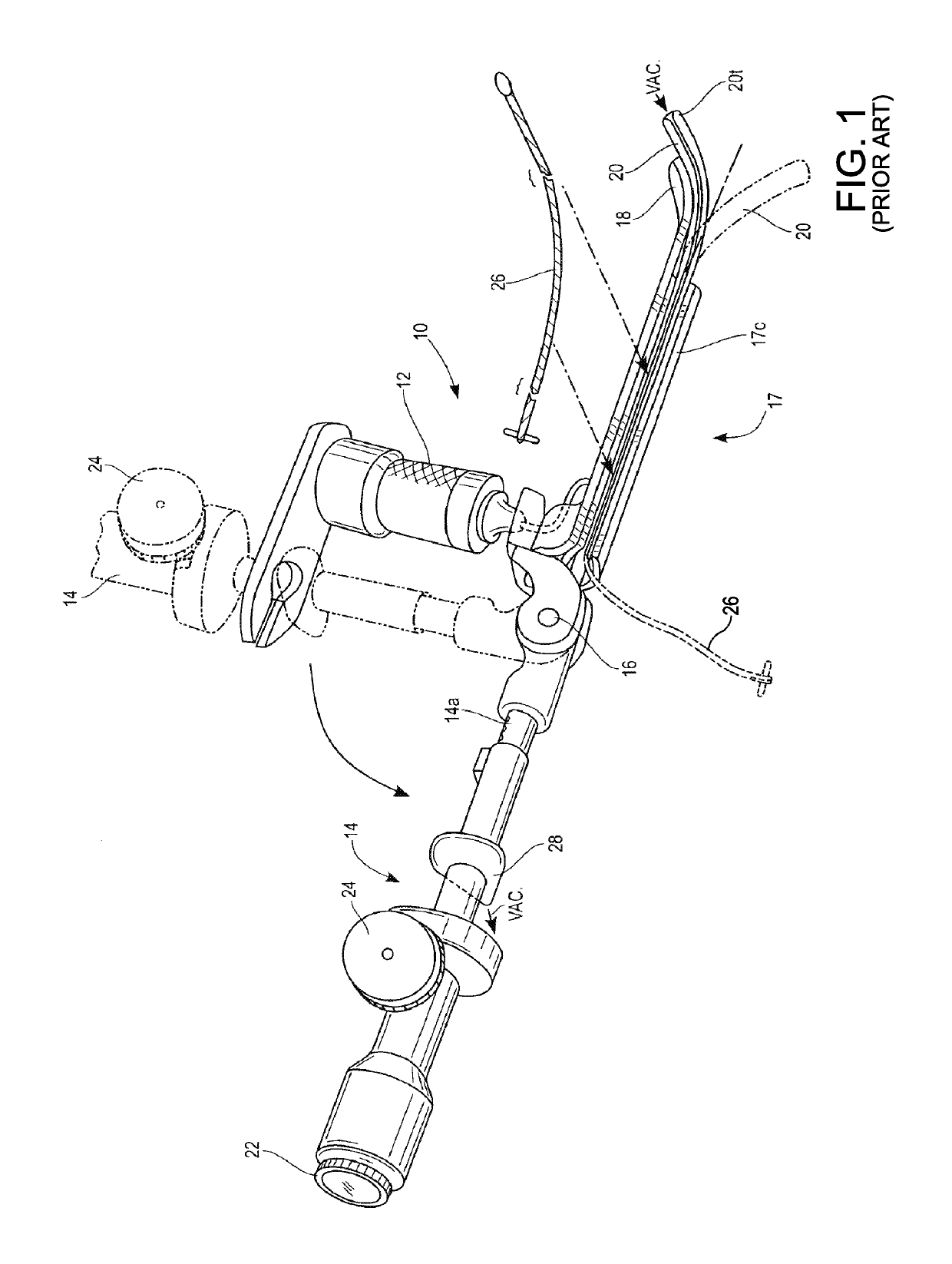
Endotracheal tube insertion device
a technology of endotracheal tube and insertion device, which is applied in the field of endotracheal tube insertion device, can solve the problems of difficult vocal cord visualization, inability to meet one or more of the requirements of available intubation instruments, and permanent patient disability and even death, and achieve the effect of improving the structure of the devi
- Summary
- Abstract
- Description
- Claims
- Application Information
AI Technical Summary
Benefits of technology
Problems solved by technology
Method used
Image

Examples
second embodiment
[0103]Referring to FIG. 11, the blade assembly is shown at 42 attached to the handle 32. The blade assembly 42 has an elongated and upwardly curved blade body 44 attached to a channel member 46. The blade body 44 includes a first or distal end 44a, and a second or proximal end 44b attached to the handle 32. Like the blade body 35, the blade body 44 has an arcuate cross-sectional shape.
[0104]The channel member 46 is attached to a first side 44c (lower side when viewing FIG. 11) of the blade body 44, includes a first portion 46a and a second portion 46b, and defines a longitudinally extending channel 46c. The channel member 46 is substantially C-shaped when viewed in cross-section and defines an elongated slot 46d that provides access to the channel 46c. Alternatively, the channel member 46 may have any desired cross-sectional shape, such as substantially oval, and substantially rectangular. The second portion 46b of the channel member 46 extends beyond the proximal end 44b of the bla...
third embodiment
[0137]the intubation assembly rod is shown at 108 in FIG. 12. The distal end 108a of the rod 108 is tapered or substantially cone-shaped and defines a leading end of the rod 108. The rod 108 includes a guide sleeve 110. The guide sleeve 110 includes a first or distal end 110a, a second or proximal end 110b, and has a longitudinally extending substantially cylindrical channel 110c formed therethrough. The guide sleeve 110 is attached to the rod 108 by a substantially flat bridge 112 that extends between the rod 108 and the guide sleeve 110. The rod 108 is otherwise substantially the same as the rod 102. Like the bridge 80, the bridge 112 may be located at any desired portion of the rod 108. The bridge 112 may have any width and length, and may be attached to the guide sleeve 110 at any point proximal to the distal end 110a of the guide sleeve 110. Further, the guide sleeve 110 may be attached to the rod 108 at any other longitudinal location or at any other desired distance from the ...
fourth embodiment
[0140]the intubation assembly rod is shown at 116 in FIG. 13. The distal end 116a of the rod 116 is tapered or substantially cone-shaped and defines a leading end of the rod 116. The rod 116 includes a guide sleeve 118. The guide sleeve 118 includes a first or distal end 118a, a second or proximal end 118b, and has a longitudinally extending substantially cylindrical channel 118c formed therethrough. The guide sleeve 118 is attached to the rod 116 by the substantially flat bridge 112 that extends between the rod 116 and the guide sleeve 118. The illustrated guide sleeve 118 also includes a longitudinally extending slot 120 formed therethrough. The rod 116 is otherwise substantially the same as the rod 102.
PUM
 Abstract
Description
Claims
Application Information