Oncolytic herpes simplex virus infected cells
a technology of infected cells, which is applied in the field of monocytes, monocyte derived cells or macrophages infected with an oncolytic herpes simplex virus, and can solve problems such as increased hypoxia
- Summary
- Abstract
- Description
- Claims
- Application Information
AI Technical Summary
Benefits of technology
Problems solved by technology
Method used
Image
Examples
example 1
HSV1716 and Human Primary Macrophages
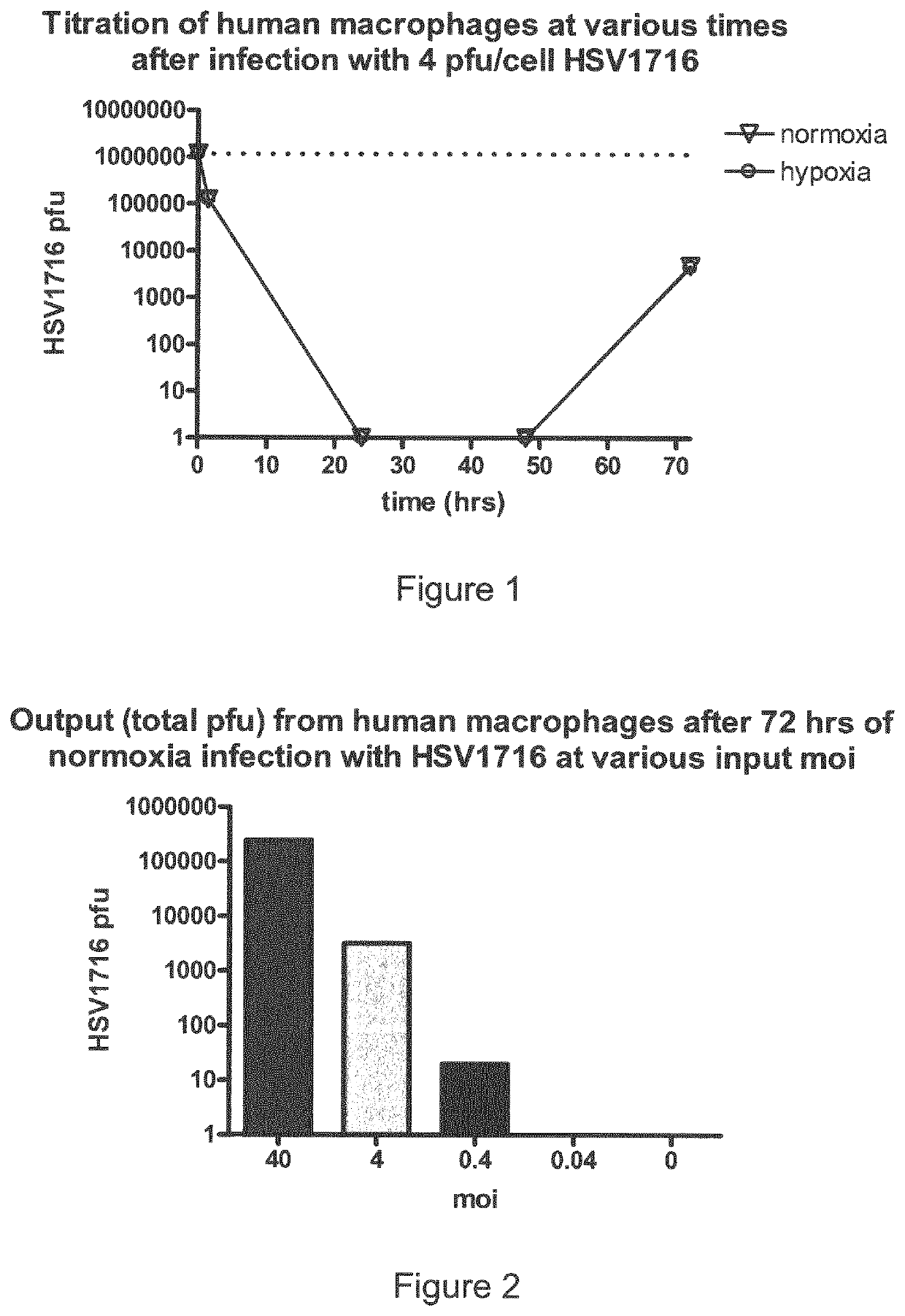
[0174]1) In an initial study human macrophages were infected with HSV1716 at approximately 4 pfu / cell and the cells were then incubated under normal and hypoxic conditions. Samples were removed at various time points after infection (+1.5 hr, +24 hrs, +48 hrs and +72 hrs) and titrated (FIG. 1).
[0175]Within 1 hour 90% of the virus had been adsorbed by the macrophages and then no virus was detectable at 24 or 48 hrs in either normoxia or hypoxia (detection limit of titration is 100 pfu / ml).
[0176]Significantly, virus was detectable at 72 hrs but the amounts at this time were similar in the normoxic vs hypoxic macrophages. This emergent virus is of significant interest as it could either be the original input which had entered some transient latent state or represent the first wave of replication in the macrophages.
[0177]2) Macrophages were infected with decreasing HSV1716 moi (40, 4, 0.4 and 0.04) and samples were titrated after 72 hrs only. Virus w...
example 2
REFERENCES FOR EXAMPLE 2
[0209]Arenberg, D. A., M. P. Keane, B. DiGiovine, S. L. Kunkel, S. R. B. Strom et al., 2000 Macrophage infiltration in human non-small-cell lung cancer: the role of CC chemokines. Cancer Immunology Immunotherapy 49: 63-70.[0210]Behnes, C. L., F. Bremmer, B. Hemmerlein, A. Strauss, P. Strobel et al., 2014 Tumor-associated macrophages are involved in tumor progression in papillary renal cell carcinoma. Virchows Archiv 464: 191-196.[0211]Brown, L. F., B. Berse, R. W. Jackman, K. Tognazzi, A. J. Guidi et al., 1995 EXPRESSION OF VASCULAR-PERMEABILITY FACTOR (VASCULAR ENDOTHELIAL GROWTH-FACTOR) AND ITS RECEPTORS IN BREAST-CANCER. Human Pathology 26: 86-91.[0212]Burke, B., 2003 Macrophages as novel cellular vehicles for gene therapy. Expert Opinion on Biological Therapy 3: 919-924.[0213]Burke, B., S. Sumner, N. Maitland and C. E. Lewis, 2002 Macrophages in gene therapy: cellular delivery vehicles and in vivo targets. Journal of Leukocyte Biology 72: 417-428.[0214]Ch...
example 3
REFERENCES FOR EXAMPLE 3
[0289]1 Culme-Seymour, E. J., Davie, N. L., Brindley, D. A., Edwards-Parton, S. & Mason, C. A decade of cell therapy clinical trials (2000-2010). Regenerative medicine 7, 455-462, doi:10.2217 / rme.12.45 (2012).[0290]2 Menasche, P. Cardiac cell therapy: lessons from clinical trials. Journal of molecular and cellular cardiology 50, 258-265, doi:10.1016 / j.yjmcc.2010.06.010 (2011).[0291]3 Fischbach, M. A., Bluestone, J. A. & Lim, W. A. Cell-based therapeutics: the next pillar of medicine. Sci Transl Med 5, 179ps177, doi:10.1126 / scitranslmed.3005568 (2013).[0292]4 Syed, B. A. & Evans, J. B. Stem cell therapy market. Nature reviews. Drug discovery 12, 185-186, doi:10.1038 / nrd3953 (2013).[0293]5 Kandalaft, L. E., Powell, D. J., Jr. & Coukos, G. A phase I clinical trial of adoptive transfer of folate receptor-alpha redirected autologous T cells for recurrent ovarian cancer. J Transl Med 10, 157, doi:10.1186 / 1479-5876-10-157 1479-5876-10-157 [pii] (2012).[0294]6 Kersha...
PUM
| Property | Measurement | Unit |
|---|---|---|
| concentration | aaaaa | aaaaa |
| concentration | aaaaa | aaaaa |
| concentration | aaaaa | aaaaa |
Abstract
Description
Claims
Application Information
 - R&D
- Intellectual Property
- Life Sciences
- Materials
- Tech Scout
- Unparalleled Data Quality
- Higher Quality Content
- 60% Fewer Hallucinations
Browse by: Latest US Patents, China's latest patents, Technical Efficacy Thesaurus, Application Domain, Technology Topic, Popular Technical Reports.
© 2025 PatSnap. All rights reserved.Legal|Privacy policy|Modern Slavery Act Transparency Statement|Sitemap|About US| Contact US: help@patsnap.com


