Compositions and methods to improve nanoparticle distribution within the brain interstitium

a brain interstitium and nanoparticle technology, applied in the direction of drug compositions, microcapsules, inorganic non-active ingredients, etc., can solve the problems of reducing the ability of nanoparticles to reach the target cells, unable to achieve therapeutically favorable distribution, etc., to reduce the adhesion of nanoparticles, improve and enhance the distribution of nanoparticles

Active Publication Date: 2020-04-28
THE JOHN HOPKINS UNIV SCHOOL OF MEDICINE
View PDF3 Cites 0 Cited by
  • Summary
  • Abstract
  • Description
  • Claims
  • Application Information

AI Technical Summary

Benefits of technology

[0012]Compositions and methods for improved distribution of nanoparticles in brain interstitium have been developed. The compositions contain nanoparticles in a hyperosmolar solution. The nanoparticle contains a first polymer, and a second hydrophilic, neutrally charged polymer that is linked to the first polymer to form a conjugate. In a preferred embodiment, the first polymer is poly(lactide-co-glycolic acid). The surface of the nanoparticle is coated with a dense coating of the hydrophilic, neutrally charged polymer, or other coating agent. The hydrophilic or neutrally charged polymer preferably includes polyethylene glycol (“PEG”), poloxomers (polyethylene oxide block copolymers), polysorbate 80 or combinations thereof. In a preferred embodiment, the dense coating is PEG. The dense coating enhances the distribution of the nanoparticles within brain interstitium by decreasing the adhesion of the nanoparticles with components of the brain extracellular matrix (ECM). The nanoparticles are preferably less than 200 nm, more preferably less than 150, most preferably less than 100 nm. In the examples, the nanoparticles have a diameter of less than or equal to 114 nm, 80 nm or 60 nm.
[0013]In some embodiments the PEG is branched. Branching enhances the density of the polyethylene glycol conjugated to the polymer. In some embodiments the polyethylene glycol has a molecular weight between 1,000 Daltons and 10,000 Daltons, such as 5,000 Daltons.
[0014]The hyperosmolar solution is formed from a solution such as saline solutions, and mannitol solutions. The hyperosmolar solution enhances the distribution of the nanoparticles within brain interstitium by increasing the pore sizes within the ECM of the brain, thereby facilitating the diffusion of the nanoparticles within the brain interstitium. In preferred embodiments, the hyperosmolar solution is saline. In a more preferred embodiment, the hyperosmolar solution is a 3% solution of saline. The examples show that the combined reduction of adhesion and increase in the pore sizes within the ECM improves the distribution of the nanoparticles away from the point of administration, and allows the escape of the nanoparticles form the perivascular spaces of the brain.

Problems solved by technology

However, recent advances in technological imaging have determined that administering a nanoparticle (NP) using CED still fails to achieve therapeutically favorable distribution (Krauze, et al., Exp Neurol, 2005, 196(1), 104-11).
Moreover, the preferable flow of NP through, and subsequent confinement within, the low resistance, fluid-filled PVS reduces their ability to reach the target cells (Krauze, et al., Exp Neurol, 2005, 196(1), 104-11; Salegio, et al., Front Neuroanat, 2014, 8, 9).

Method used


Image

  • Compositions and methods to improve nanoparticle distribution within the brain interstitium
  • Compositions and methods to improve nanoparticle distribution within the brain interstitium
  • Compositions and methods to improve nanoparticle distribution within the brain interstitium

Examples


example 1

f Coating Particles with Non-adhesive Coating

[0141]Materials and Methods

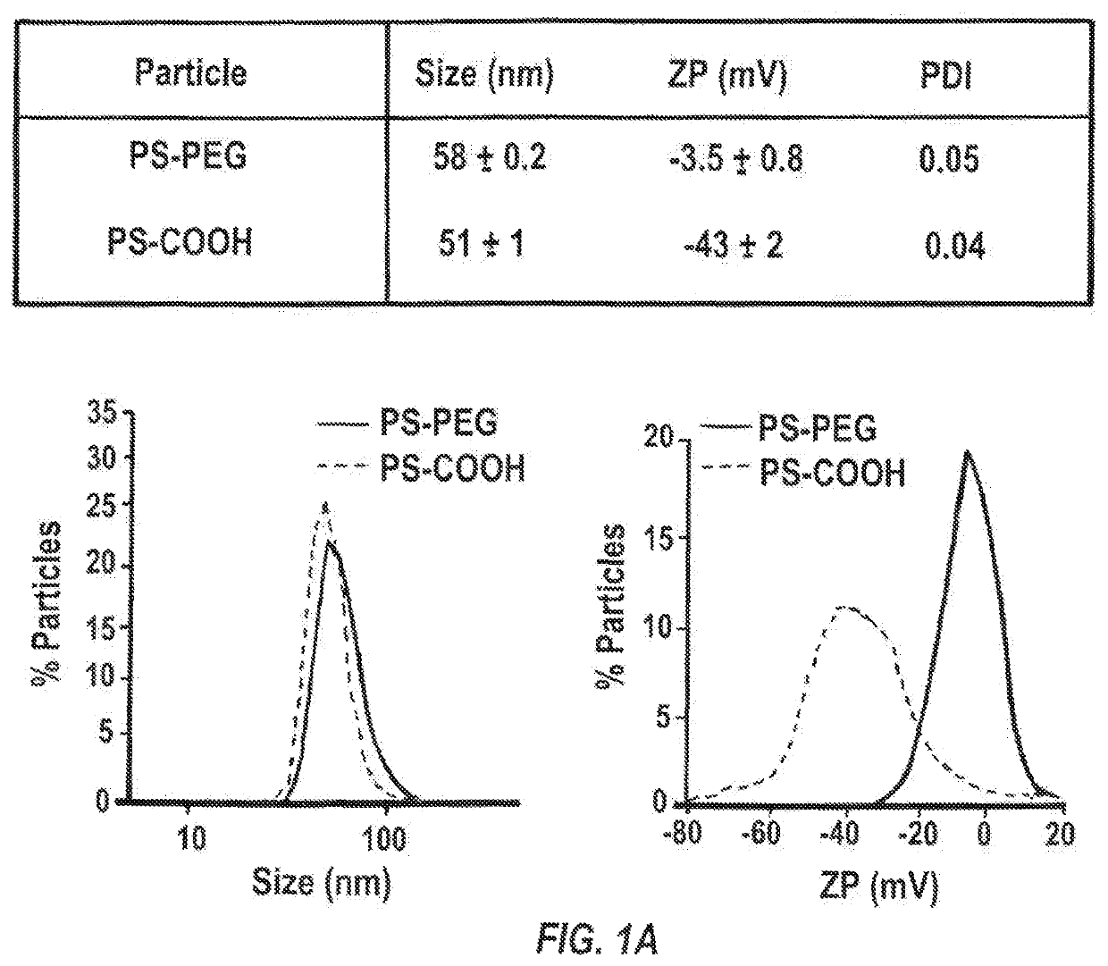
[0142]i. Nanoparticle Preparation and Characterization

[0143]40-nm dark red fluorescent carboxylated polystyrene microspheres (PS-COOH) (Life Technologies, Grand Island, N.Y.) were modified by conjugating a dense layer of 5 kDa methoxy-PEG-amine (Creative PEGworks, Winston Salem, N.C.), onto the surface, according to a previously published protocol (Nance, E. A., et al., Sci Transl Med, 2012, 4(149), 149ra119), to obtain densely PEGylated polystyrene nanoparticles (PS-PEG). PLGA (75:25) (MW: 15 kDa; Jinan Daigang Biomaterials Co. Ltd., Jinan, China) and PLGA-PEG (75:25) (25 wt % PEG; Jinan Daigang Biomaterials Co. Ltd., Jinan, China) nanoparticles were formulated using the single emulsion process according to a previously published protocol (Nance, E., et al., ACS Nano, 2014, 8(10), 10655-10664). Briefly, PLGA-PEG and PLGA polymer were fluorescently labeled with AlexFluor 647 and AlexaFluor 555 cadaverine dye (Mo...

example 2

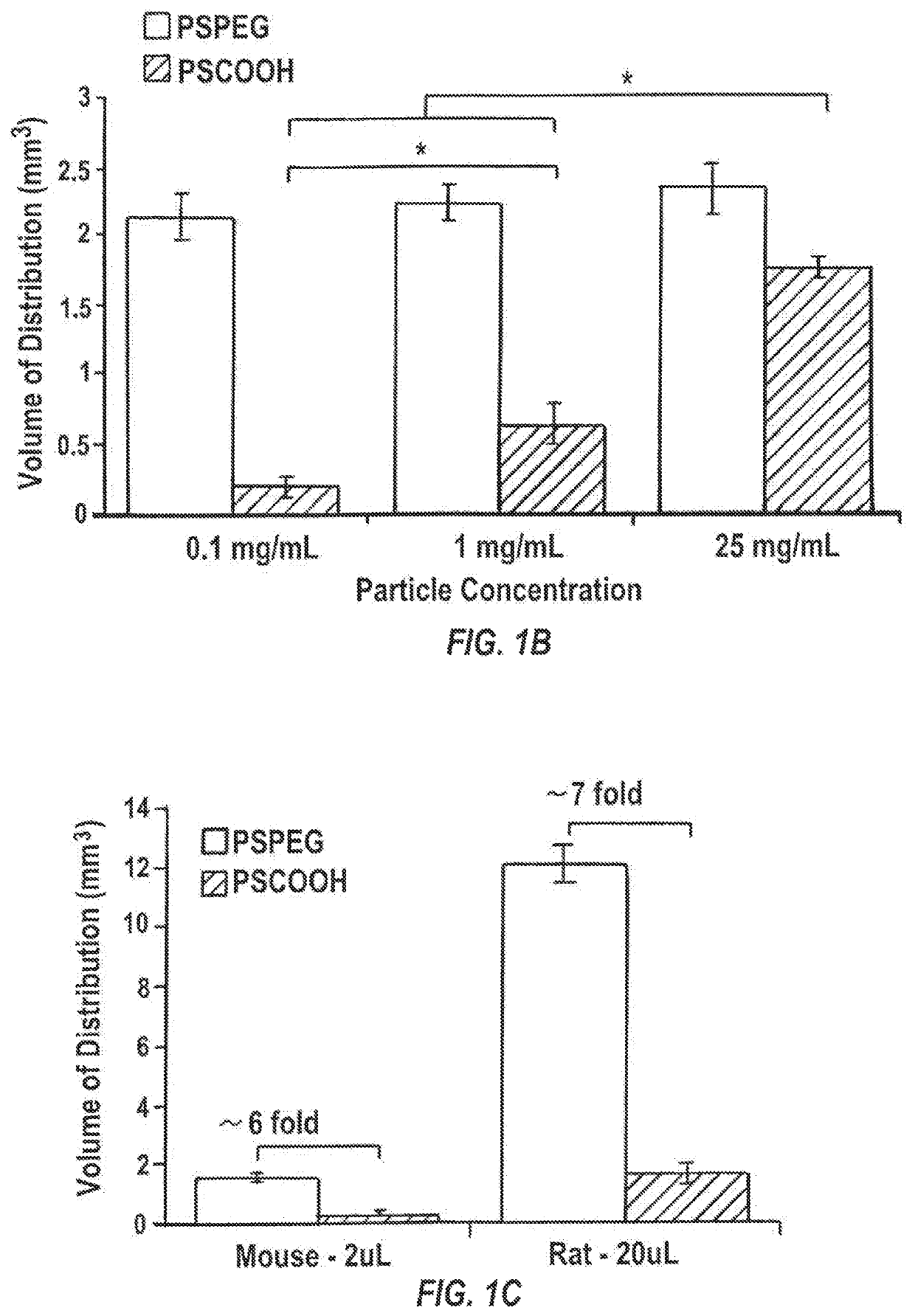
f Different Concentrations of Infusate Composition

[0159]Materials and Methods

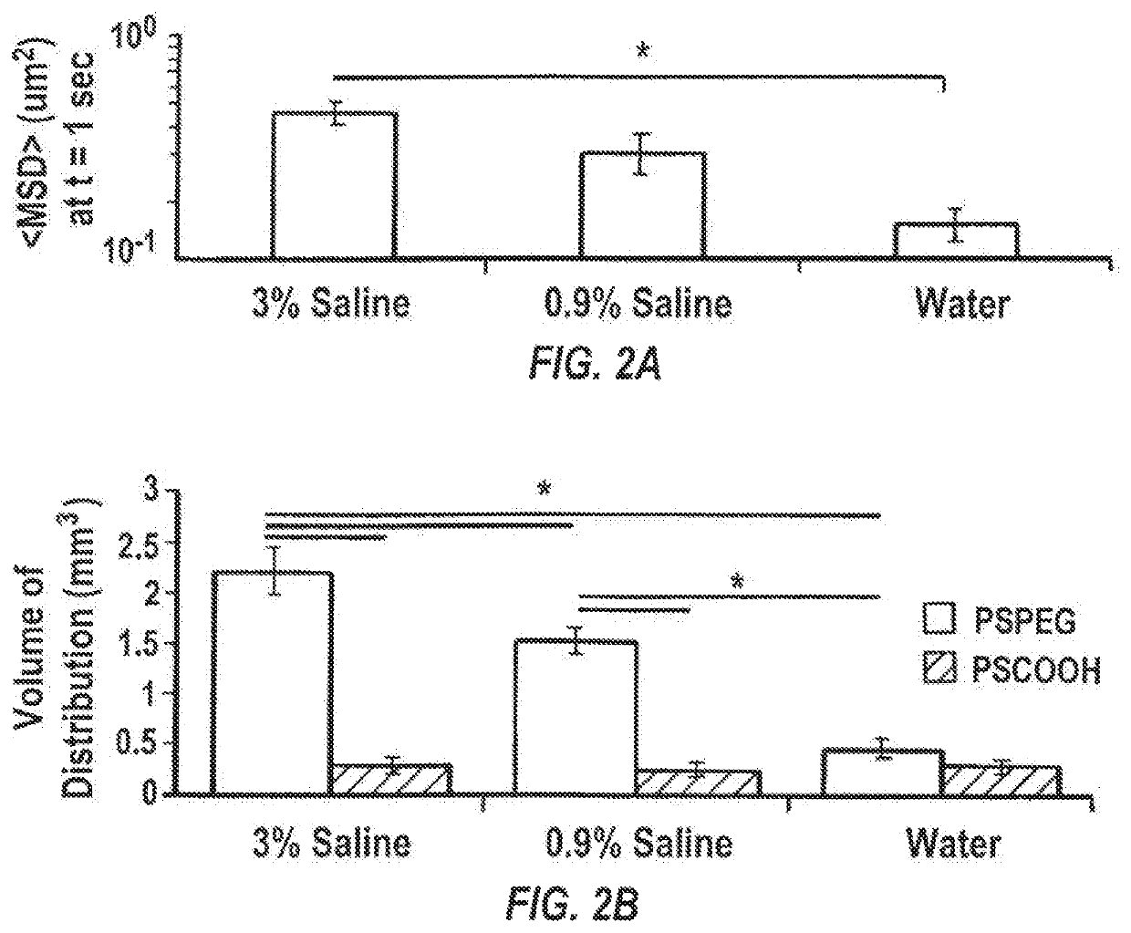
[0160]i. Ex Vivo Characterization of Brain Pore Sizes

[0161]Brains from female CF-1 mice were harvested and multiple particle tracking was conducted on nanoparticles injected into 1.5 mm thick brain slices according to a slightly modified protocol of Nance, et al., Sci Transl Med, 2012, 4(149), 149ra119. Briefly, the harvested rodent brain was rinsed in chilled artificial spinal fluid and sliced at 1.5 mm intervals using a Zivic mouse brain mold (Zivic Instruments, Pittsburgh, Pa.). Individual brain slices were immersed in infusate solutions (water, 0.9% saline, 3% saline, 10% mannitol, or 25% mannitol) for 5 minutes. Brain slices were removed and mounted on a custom made well and 0.5 μL of fluorescently labeled PS-PEG nanoparticles were injected into the cortex. A coverslip was glued on top of the specimen to prevent bulk flow in the tissue. The particle trajectories were recorded as 20 second movies at an ...


PUM

PropertyMeasurementUnit
diameteraaaaaaaaaa
diameteraaaaaaaaaa
diameteraaaaaaaaaa

Abstract

Improved distribution can be achieved by delivering nanoparticles possessing non-adhesive surfaces via CED in a hyperosmolar infusate solution. This delivery strategy minimizes the hindrances imposed by the brain extracellular matrix and reduces the concentration of therapeutic that is confined within perivascular spaces.

Description

CROSS-REFERENCE TO RELATED APPLICATIONS[0001]This application is a National Phase application under 35 U.S.C. 371 of PCT / US2016 / 050967, filed Sep. 9, 2016, which claims priority to and benefit of U.S. Provisional Application No. 62,217,194, filed Sep. 11, 2015, which is hereby incorporated herein by reference in its entirety.STATEMENT REGARDING FEDERALLY SPONSORED RESEARCH OR DEVELOPMENT[0002]This invention was made with government support under grant CA164789 awarded by the National Institutes of Health. The government has certain rights in the invention.FIELD OF THE INVENTION[0003]This invention is generally in the field of compositions and methods for the enhanced delivery of therapeutic, diagnostic, or prophylactic agents, and in particular, the delivering of these agents into the brain by facilitating their escape from perivascular spaces in order to achieve widespread volumes of distribution of these agents in brain interstitium.BACKGROUND OF THE INVENTION[0004]Many brain dise...

Claims


Application Information

Patent Timeline
Patent Type & Authority Patents(United States)
IPC IPC(8): A61K9/14A61K49/00A61K9/00A61K9/51A61K47/02A61P25/00
CPCA61K9/0085A61K47/02A61K9/5138A61K9/5115A61K9/5153A61K49/0032A61K49/0093A61K9/5146A61K9/5123A61P25/00
Inventor ZHANG, CLARKMASTORAKOS, PANAGIOTISSUK, JUNG SOOHANES, JUSTIN
Owner THE JOHN HOPKINS UNIV SCHOOL OF MEDICINE
Why Eureka
Try Eureka