Apparatus for vessel characterization

a technology of apparatus and stenosis, applied in the field of apparatus, system and method of stenosis of vessels and vessel modeling, can solve the problems of obstructed visual evaluation of the stenotic region, unable to accurately estimate the cross-sectional shape of the stenosis, etc., and achieve the effect of simple construction

Active Publication Date: 2020-06-30
KONINKLJIJKE PHILIPS NV
View PDF10 Cites 0 Cited by
  • Summary
  • Abstract
  • Description
  • Claims
  • Application Information

AI Technical Summary

Benefits of technology

[0009]The apparatus is configured to derive the cross sectional area of the vessel based on two pressure measurements at two distinct locations within the vessel, without needing to use information from imaging techniques such as fluoroscopy in conjunction with radio-opaque contrast agents.
[0010]In an embodiment, the apparatus is further configured to receive from the instrument within the vessel a third pressure measurement signal at a third measurement location and to derive a second cross sectional area for a second reference position of the vessel based on the third pressure measurement signal and one of the first and second pressure measurement signals. The benefit of deriving multiple cross sectional areas at multiple reference positions along the vessel is that the condition of the vessel can be evaluated based on the change of the cross sectional diameters at the respective reference positions. In such a way detection of potential narrowing or widening of the vessel is possible without using imaging techniques.
[0011]In an embodiment, the apparatus is further configured to render a representation of the vessel based on the first and second cross sectional areas and the first and second reference positions. A graphical representation based on the distance between the two reference positions and the cross sectional areas at the respective locations improves the characterization of the vessel by allowing a quick visual interpretation of the condition of the vessel.
[0012]In a further embodiment, the first, second and third pressure measurement signals are temporal measurement signals of a pulsatile blood flow within the vessel; the first and second cross sectional areas are derived for subsequent phases of the pulsatile blood flow; and the representation of the vessel comprises a sequence of representations of the vessel corresponding to the subsequent phases of the pulsatile blood flow. The pulsatile blood flow is caused by the cardiac heart beat cycle. The graphical representation of the vessel depending on the phases of the pulsatile blood flow allows an improved evaluation of potential conditions of the vessel where elasticity of the vessel segments are influenced by the magnitude of the blood pressure. Such conditions may be an asymmetric narrowing of the vessel, formation of aneurysm, etc.
[0013]In an embodiment, the apparatus is further configured: to receive morphological information of the vessel from an imaging unit; to render a composite representation comprising: a morphological representation of the vessel based on the morphological information and the representation of the vessel based on the first and second cross sectional areas and the first and second reference positions. The representation based on morphological information received from the imaging unit enriches the information available for evaluation of the condition of the vessel, providing the physician with additional information on the orientation of the vessels within the body of the patient.
[0016]In an embodiment, the system is further configured to receive morphological information of the vessel from an imaging unit; to render a composite representation comprising: a morphological representation of the vessel based on the morphological information and the representation of the vessel based on the first and second cross sectional areas and the first and second reference positions. A combination of the representation of the vessel based on pressure measurements and the representation based on morphological information enriches the possibilities for evaluation of the vessel condition. An overlay of the two representations may be based on visual interpretation by the physician of particular features (narrowing, widening, bends, etc.) of the vessel from the two representations, and it may be enabled for the physician by a drag and drop technique applied for one of the representations. Alternatively, the overlay may be based on automatic registration by a computer vision algorithm. Once the two representations are registered the measurement locations are also known within the morphological representation. This would replace the need for device tracking, while the intravascular pressure data can be displayed at the correct location within the morphological representation.

Problems solved by technology

The weakness of the technique is that the cross-sectional shape of the stenosis can only be roughly estimated even by using multiple projections.
Furthermore, such a visual evaluation of the stenotic region may be obstructed by other vessels that overlap the stenosis in the image, as well as by image artefacts and noise.

Method used


Image

  • Apparatus for vessel characterization
  • Apparatus for vessel characterization
  • Apparatus for vessel characterization

Examples


Embodiment Construction

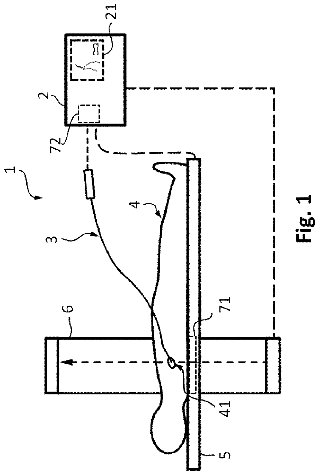
[0042]In FIG. 1 an embodiment of the system 1 for characterization of a vessel of a living being 4 is presented, wherein the living being may be a person or an animal. The system 1 comprises an apparatus 2 and an instrument 3, wherein the instrument is configured to provide pressure measurement signals from within a vessel of a portion 41 of the circulatory system of the patient 4, and the apparatus 2 is configured to ascertain characteristics of the vessel based on the pressure measurements. The apparatus 2 comprises a display 21 for rendering the characteristics of the vessel. Alternatively, the apparatus may be configured to provide signals to an external display on which the designated characteristics are presented to the physicians.

[0043]In an embodiment, the system comprises an imaging unit 6 for acquiring two-dimensional or three-dimensional morphological information of the vessel structure of interest upon injection of a contrast agent bolus into the targeted vasculature. Th...


PUM


Abstract

The invention discloses an apparatus (2), a system (1) and a method (100) for characterization of vessels and for vessel modeling. The cross sectional area (A1) of the vessel is derived from pressure measurements (p1, p2) obtained by an instrument (3) from within the vessel. When multiple cross sectional areas (A1, A2) are derived for multiple reference positions (r1, r2) based on pressure measurements (p1, p2, p3) along the vessel, a representation (20, 30) of the vessel can be rendered, without requiring any imaging modality. Furthermore, the effect of the pulsatile blood flow on the elasticity of the vessel walls can be visualized, supporting assessment of a stenosis or an aneurysm formation along the vessel.

Description

CROSS-REFERENCE TO PRIOR APPLICATIONS[0001]This application is the U.S. National Phase application under 35 U.S.C. § 371 of International Application No. PCT / EP2017 / 054539, filed on Feb. 28, 2017, which claims the benefit of European Patent Application No. 16158610.2, filed on Mar. 4, 2016. These applications are hereby incorporated by reference herein.FIELD OF THE INVENTION[0002]The invention relates to an apparatus, system and method for characterization of vessels and for vessel modeling.BACKGROUND OF THE INVENTION[0003]A typical technique for identifying stenotic regions in a blood vessel is using angiography, whereas the functional impact of the stenosis is quantified with the fractional flow reserve (FFR) technique. FFR is calculated from the ratio of pressures at the distal and proximal ends of the stenosis, measured with a sensor positioned at the tip of a wire. The FFR value is a measure of the severity of the stenosis, and it can be established whether the stenosis limits ...

Claims


Application Information

Patent Timeline
Patent Type & Authority Patents(United States)
IPC IPC(8): A61B5/0215A61B5/06A61B5/00G06T7/00A61B5/107A61B5/02A61B8/08A61B6/12
CPCG06T7/0012A61B5/0215A61B5/02007A61B5/0035A61B5/063A61B5/02028A61B5/1076A61B8/0841A61B5/7425A61B6/12A61B5/02158A61B5/066A61B5/064
Inventor HAASE, CHRISTIANGRASS, MICHAELSCHAEFER, DIRK
Owner KONINKLJIJKE PHILIPS NV
Why Eureka
Try Eureka