Spatially distinguished, multiplex nucleic acid analysis of biological specimens
a technology of multiplex nucleic acid and biological specimens, applied in the field of spatial distinction and multiplex nucleic acid analysis of biological specimens, can solve the problems of loss of time needed to treat an aggressive cancer, more complex mosaics within the tumor, adverse effects on patients both emotionally and physically
- Summary
- Abstract
- Description
- Claims
- Application Information
AI Technical Summary
Benefits of technology
Problems solved by technology
Method used
Image
Examples
example i
Spatially Tagging mRNA from a Tissue Sample Using Illumina Flow Cells
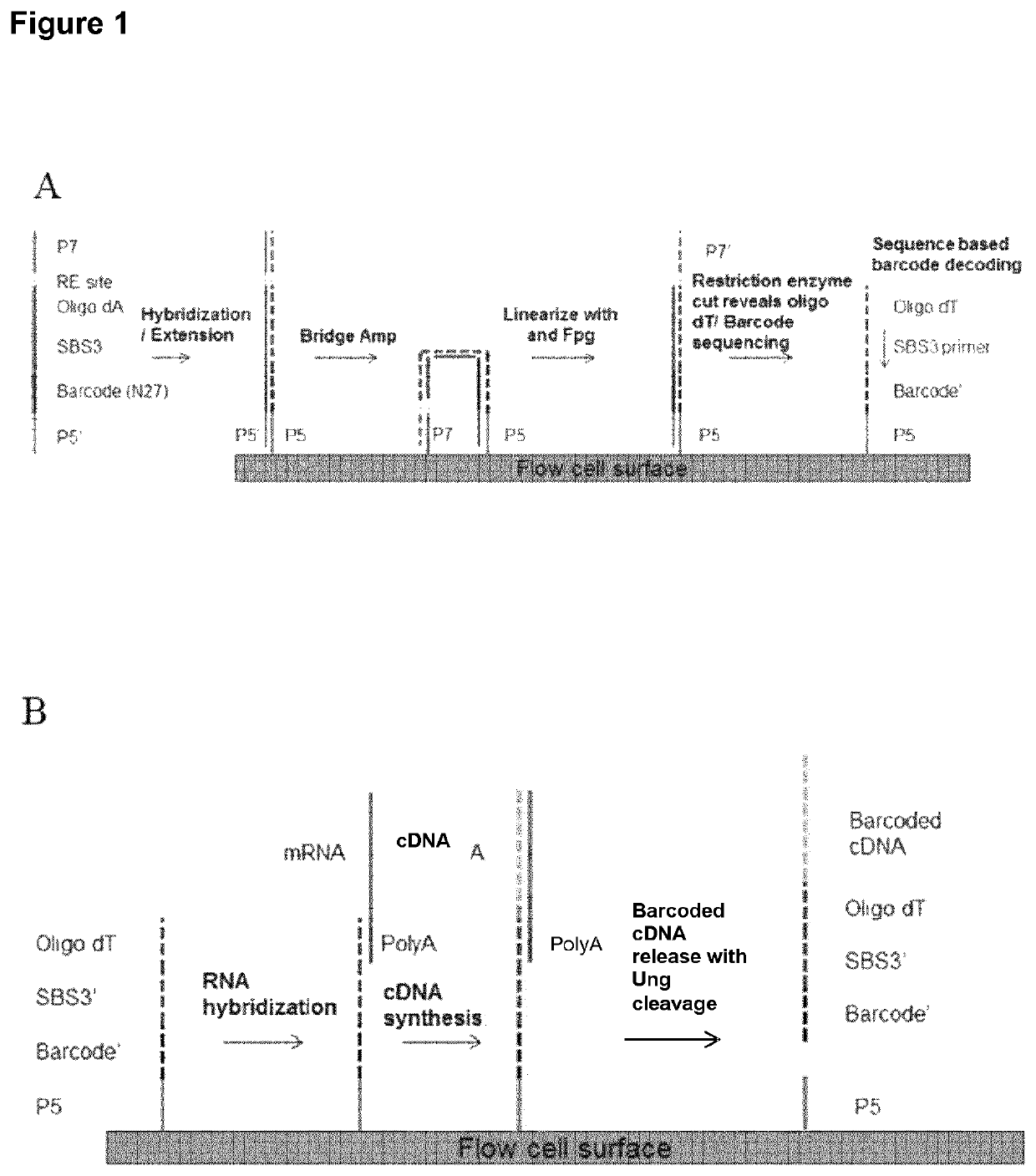
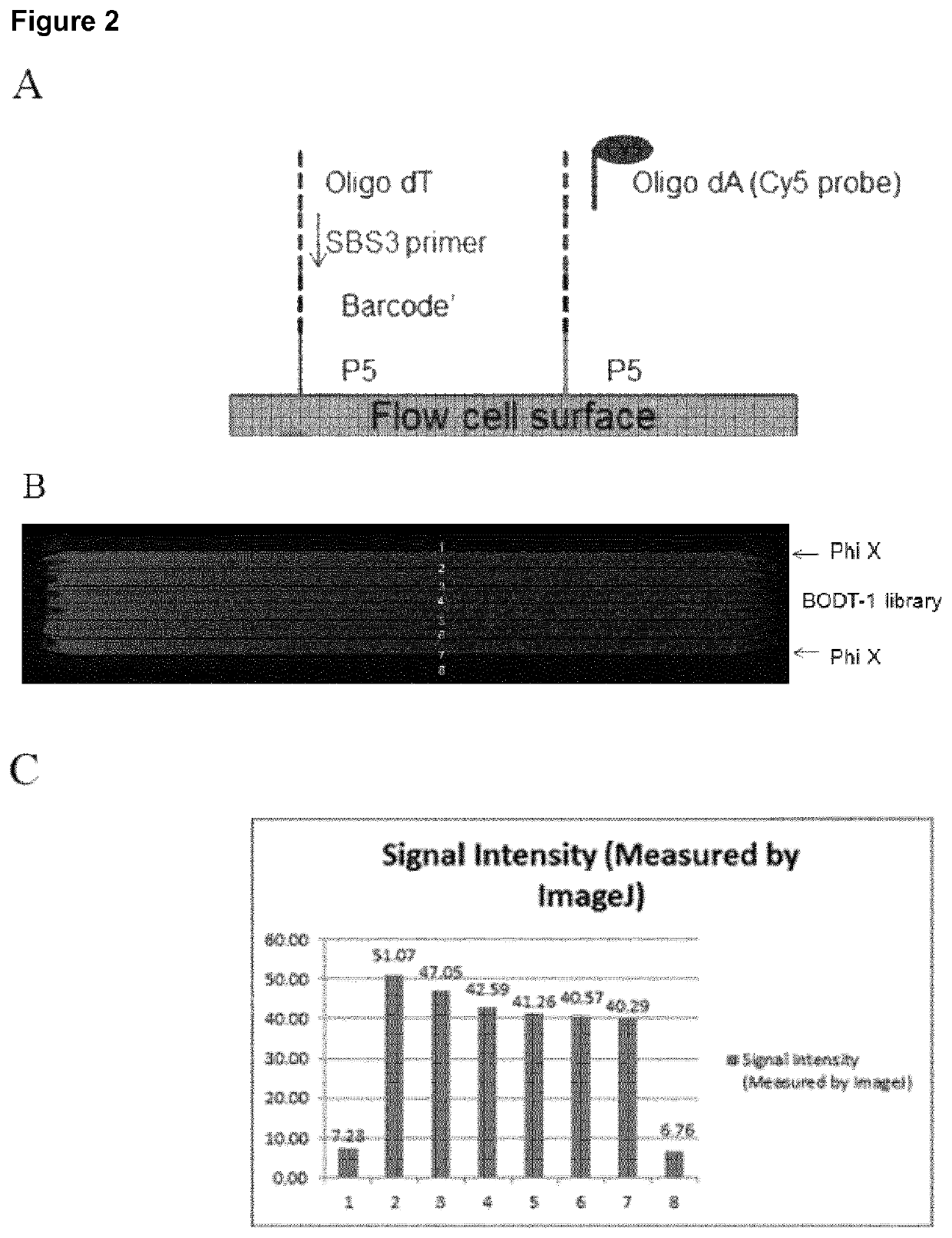
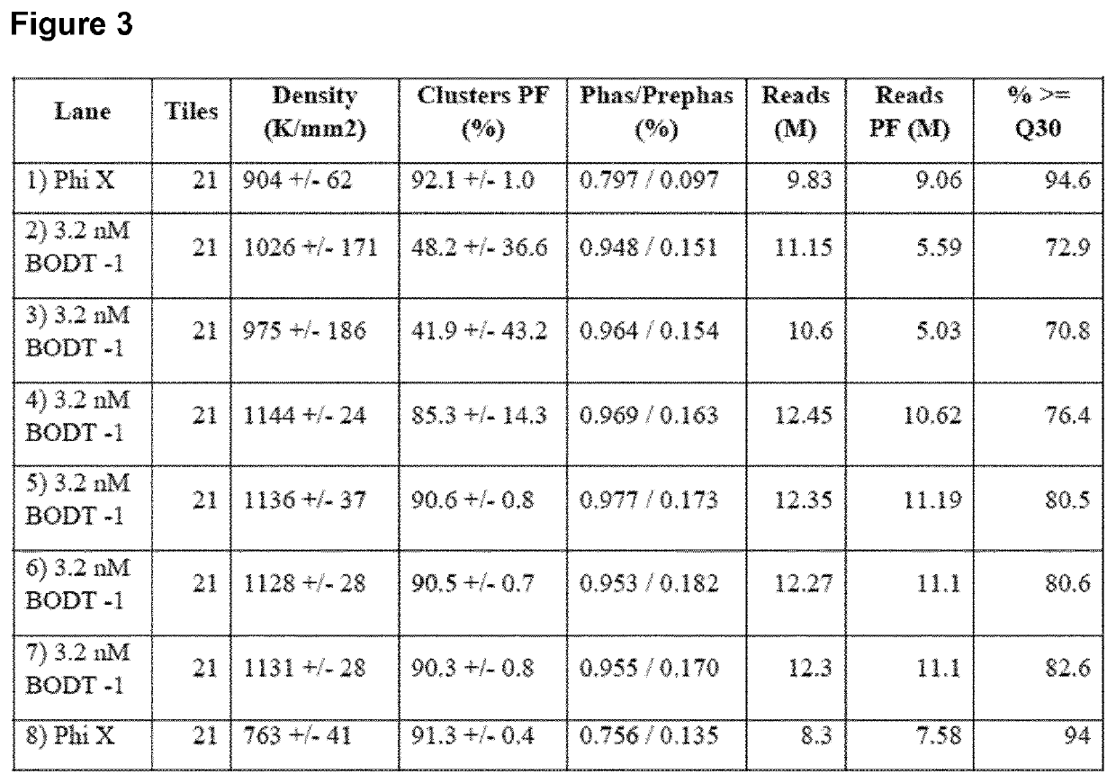
[0129]A method for generating barcoded oligo-dT containing clusters, then revealing the barcoded oligo-dT with a restriction enzyme digest followed by sequencing is described in FIG. 1. A library of fragments containing a single stranded, barcoded oligo-dA, P5′, P7, SBS3 sequencing primer binding site and a BspH1 restriction enzyme site (shown in the top panel of FIG. 1) were prepared by oligo synthesis (Integrated DNA Technologies). The barcodes were 27mers and were randomly generated during synthesis. The binding site for the SBS3 sequencing primer was included for decoding of the barcode by sequencing. An oligo-dA stretch was included to generate an oligo dT site upon clustering and linearization. Bridge amplification and clustering were performed according to standard cluster chemistry (Illumina TruSeq PE Cluster Kit v3 cBot P / N: 15037931) on an Illumina GA flow cell using manufacture's recommended protocol.
[01...
example ii
Cell Adhesion on Illumina Flow Cells
[0134]Single cells were captured on a patterned flow cell (HiSeq X10 flow cell, Illumina). All reagent flow steps were performed using a peristaltic pump or the cBOT cluster generation instrument (Illumina). Briefly, nuclease free water was flowed on all lanes of the patterned flow cell followed by 30-70K Poly D Lysine Solution (100 μg / ml and 20 μg / ml) at a flow rate of 100 μl / min for 8 min. Heat inactivated Fetal Bovine Serum (Life Technologies #10082-139) was also tested as an adhesive. The adhesives were incubated on the flow cell lanes for 1 hr, followed by a 1× PBS+0.5% Pluronic F-68 (Life Technologies #24040-032) wash. Next, the cells were adhered to the coated flow cells by flowing 5 to 50 cells / μl or approximately 100-1000 cells per lane at a rate of 100 μl / min, followed by an incubation step for 60 min to bind the cells. The flow cell was washed with 1× PBS / 0.5% pluronic at a rate of 75 μl / min. If cells were fixed on the flow cell, 1% Par...
example iii
Spatially Localized Capture of Target mRNA by Probes Attached to a Gel Surface
[0136]This example describes creation of a lawn of poly T probes on a gel coated slide, placement of tissue slices on top of a lawn of poly T probes, release of RNA from the tissue sections, capture of the released mRNA by the poly T probes, reverse transcription to Cy 3 label the poly T probes, removal of the tissue and imaging of the slide.
[0137]FIG. 7, Panel A shows a diagrammatic representation of steps and reagents used to create probes attached to a gel. Briefly, a microscope slide was coated with silane free acrylamide (SFA), P5 and P7 primers were attached (see US Pat. App. Pub. No. 2011 / 0059865 A1, which is incorporated herein by reference), probes having a poly A sequence and either a P5 or P7 complementary sequence were hybridized to the P5 and P7 primers, respectively, and the P5 and P7 primers were extended to produce poly T sequence extensions. A quality control step was performed by hybridiz...
PUM
| Property | Measurement | Unit |
|---|---|---|
| area | aaaaa | aaaaa |
| area | aaaaa | aaaaa |
| area | aaaaa | aaaaa |
Abstract
Description
Claims
Application Information
 - R&D
- Intellectual Property
- Life Sciences
- Materials
- Tech Scout
- Unparalleled Data Quality
- Higher Quality Content
- 60% Fewer Hallucinations
Browse by: Latest US Patents, China's latest patents, Technical Efficacy Thesaurus, Application Domain, Technology Topic, Popular Technical Reports.
© 2025 PatSnap. All rights reserved.Legal|Privacy policy|Modern Slavery Act Transparency Statement|Sitemap|About US| Contact US: help@patsnap.com


