
Visualization of an image object relating to an instrucment in an extracorporeal image

a technology of image object and instrument, applied in image enhancement, instruments, catheters, etc., can solve the problems of patient and staff being continuously exposed to ionizing x-ray radiation, and having a detrimental effect on the health of patients and sta

Active Publication Date: 2021-05-11
KONINKLJIJKE PHILIPS NV
View PDF32 Cites 1 Cited by
  • Summary
  • Abstract
  • Description
  • Claims
  • Application Information

AI Technical Summary

Benefits of technology

This approach provides real-time, intuitive visualization of instrument positions and orientations within the body, reducing the need for continuous x-ray imaging and minimizing radiation exposure to patients and staff.

Problems solved by technology

This also means that the patient and staff are continuously exposed to ionizing x-ray radiation in these applications.
This may have a detrimental effect on the health of the patient and staff.

Method used


Image

  • Visualization of an image object relating to an instrucment in an extracorporeal image
  • Visualization of an image object relating to an instrucment in an extracorporeal image
  • Visualization of an image object relating to an instrucment in an extracorporeal image

Examples


Embodiment Construction

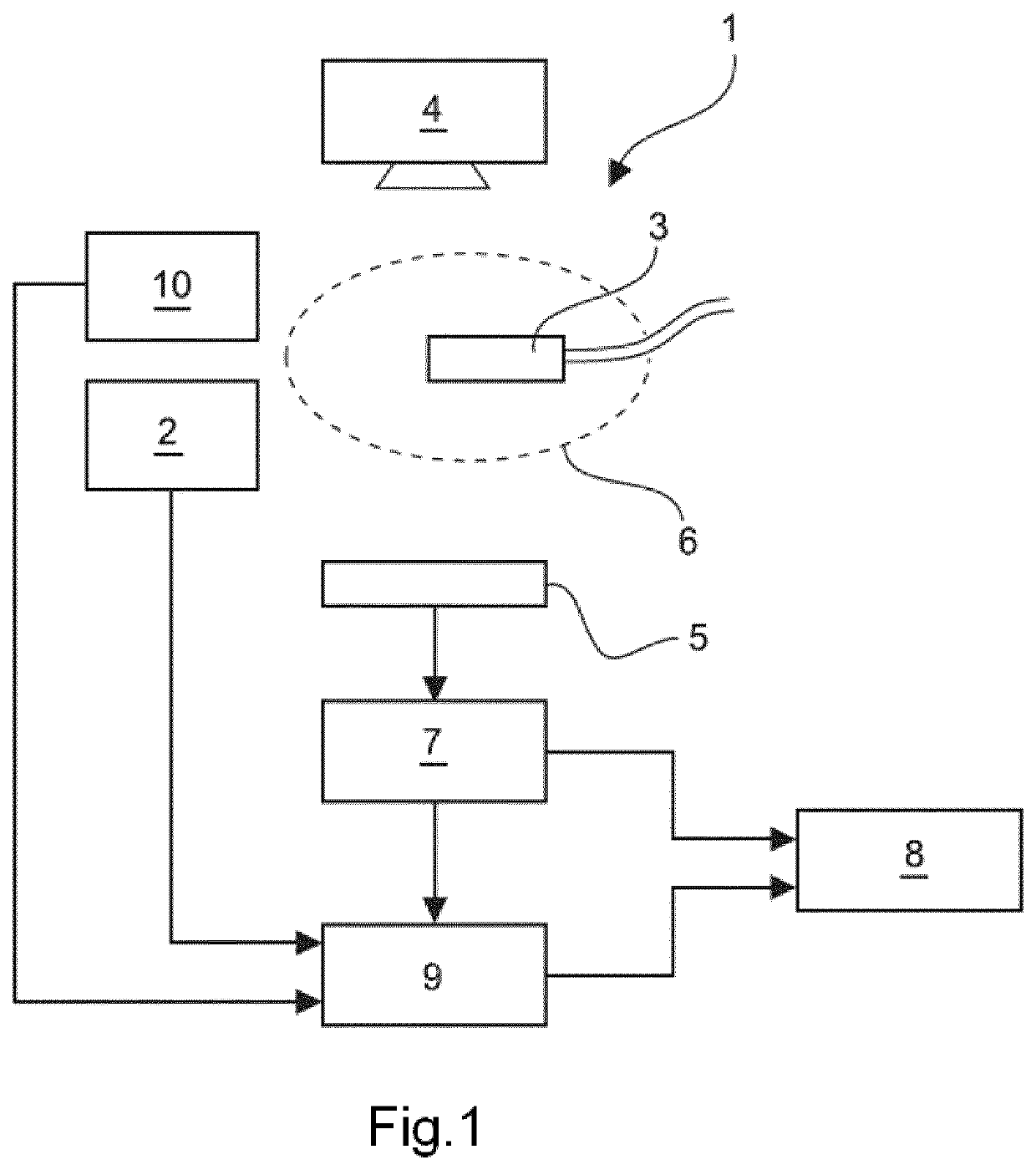
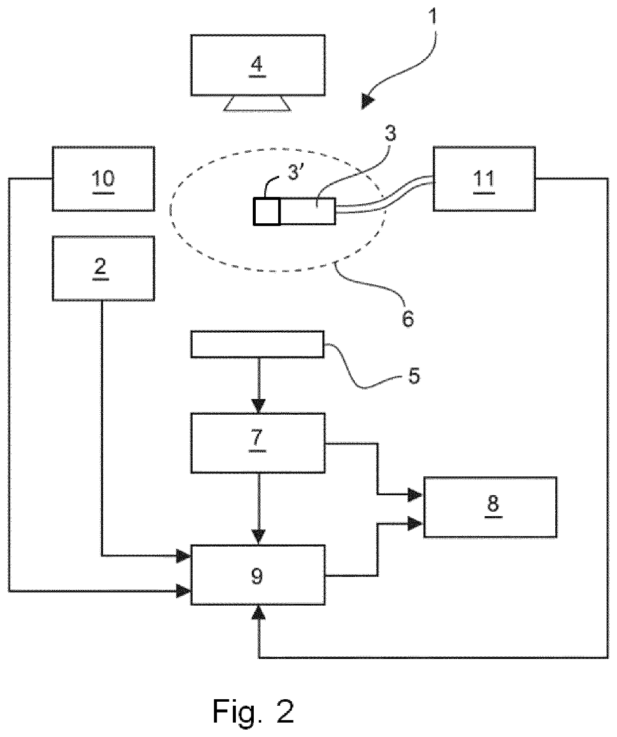
[0042]FIG. 1 schematically and exemplarily shows a first embodiment of a system comprising an extracorporeal image acquisition system 1 used in order to provide images of a region of interest within a human or animal patient body for diagnostic purposes and / or during surgical interventions (particularly minimally invasive surgical interventions). The region of interest may include the heart of the patient as it is the case in one embodiment referred to herein below. However, the system may likewise be used in order to image other body regions.

[0043]The extracorporeal image acquisition system 1 may be one of an x-ray based imaging device, a magnetic resonance imaging device or an ultrasound imaging system, capable of providing images of the interior of the body of the patient.

[0044]In the following, for elucidating the invention, the x-ray device is used as extracorporeal image acquisition system, however it can be understood by those skilled in the art that an x-ray image of a body ...


PUM


Abstract

The invention relates to an apparatus and a system for visualizing an image object relating to an instrument (3), particularly a medical instrument, in an extracorporeal image. The image object may comprise a representation of the instrument (3) or an intracorporeal image (40) acquired using the instrument (3). The system comprises an extracorporeal image acquisition device (1) for acquiring extracorporeal images with respect to an extracorporeal image frame and a tracking arrangement (2) for tracking the instrument (3) independent of the extracorporeal images with respect to a tracking frame. Further, the invention relates to a method carried out in the system.

Description

CROSS-REFERENCE TO PRIOR APPLICATIONS[0001]This application is the U.S. National Phase application under 35 U.S.C. § 371 of International Application No. PCT / EP2017 / 073379, filed on Sep. 18, 2017, which claims the benefit of European Patent Application No. 16190267.1, filed on Sep. 23, 2016. These applications are hereby incorporated by reference herein.FIELD OF THE INVENTION[0002]The invention relates to an apparatus, a system and a method for visualization of an image object relating to an instrument in an extracorporeal image. Moreover, the invention relates to a computer program for carrying out the method using a processing unit.BACKGROUND OF THE INVENTION[0003]In medical interventions, fluoroscopy is often employed in order to track the position and orientation of instruments inserted into a region of interest within a body of a patient. In fluoroscopy, x-ray images of the region of interest are (quasi-) continuously acquired so that moving x-ray images are generated which all...

Claims


Application Information

Patent Timeline
Patent Type & Authority Patents(United States)
IPC IPC(8): A61B34/00A61B8/08A61B6/00A61B6/12A61B8/00A61B34/20A61B5/00G06T7/30G06T7/70G06T7/20
CPCA61B34/25A61B6/12A61B6/5247A61B6/5288A61B8/461A61B8/5284A61B34/20G06T7/20G06T7/30G06T7/70A61B5/0084A61B2034/2051A61B2034/2055A61B2034/2063A61B2034/2065A61B2034/2074G06T2207/10101G06T2207/10116G06T2207/10132
Inventor HARKS, GODEFRIDUS ANTONIUSNIJHOF, NIELS
Owner KONINKLJIJKE PHILIPS NV
Learn More