Intracardiac ultrasound catheter handheld adapter

a technology of ultrasound catheter and handheld adapter, which is applied in the direction of ultrasonic/sonic/infrasonic image/data processing, catheters, applications, etc., can solve the problems of life-threatening, atrial esophageal fistula following af ablation can be fatal, and intravascular access can have complications

Active Publication Date: 2021-05-18
KUSUMOTO WALTER
View PDF33 Cites 2 Cited by
  • Summary
  • Abstract
  • Description
  • Claims
  • Application Information

AI Technical Summary

Benefits of technology

[0011]With this invention, a cardiac electrophysiology mapping system is utilized to visualize blood vessels for percutaneous access. Visualization of the blood vessels can be accomplished by using an intracardiac ultrasound handheld adapter in conjunction with the intracardiac ultrasound catheter (or other intravascular imaging device). This allows for visualization of the blood vessel contours through the cardiac electrophysiology mapping system. A percutaneous needle with mounted magnetic sensors and / or electrodes, as discussed herein, can allow for direct and real time visualization of vessel access on a display of the cardiac electrophysiology mapping system.

Problems solved by technology

However, complications from intravascular access and esophageal ulcers are relatively common and can be life threatening.
However, intravascular access can have complications.
Additionally, esophageal ulceration following AF ablation is a relatively common occurrence (50%), and atrial esophageal fistula following AF ablation can be fatal.
These iatrogenic complications cause significant morbidity and mortality within the hospital.
Fluoroscopy is sometimes utilized, but exposes both patient and operator to radiation.
CT and MRI guidance have been reported, however would require access to CT / MRI which may not be readily available in the cardiac catheterization lab.
Various esophageal protective strategies including esophageal temperature monitoring and physical manipulation of the esophagus have not shown to be conclusively protective of the esophagus.
However, this technique requires the operator to simultaneously hold the echo probe and percutaneous needle and the echo probe can frequently obstruct the point of access.

Method used


Image

  • Intracardiac ultrasound catheter handheld adapter
  • Intracardiac ultrasound catheter handheld adapter
  • Intracardiac ultrasound catheter handheld adapter

Examples


Embodiment Construction

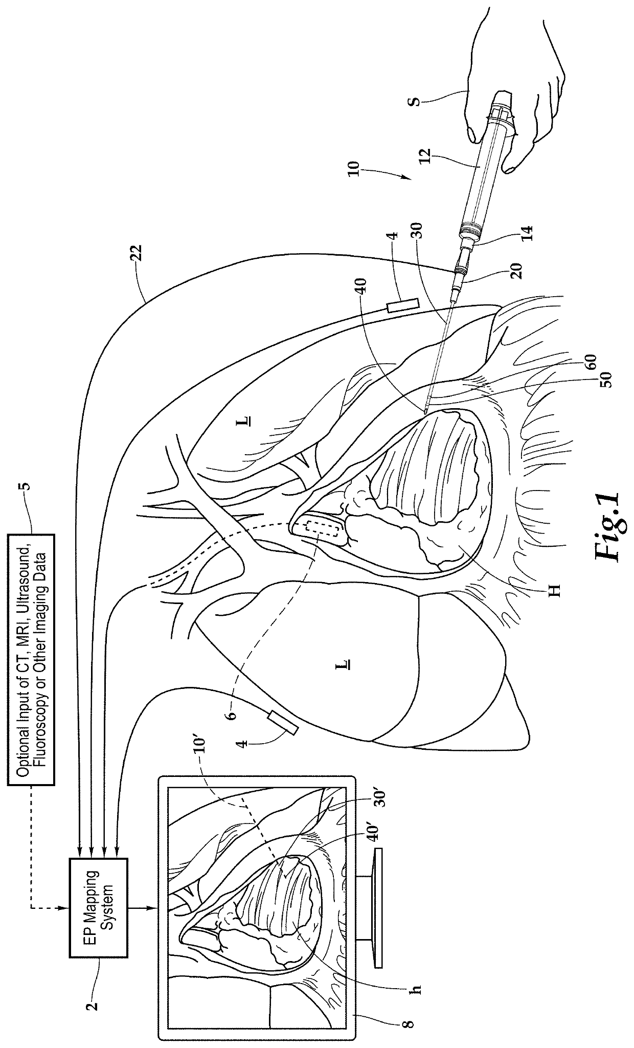
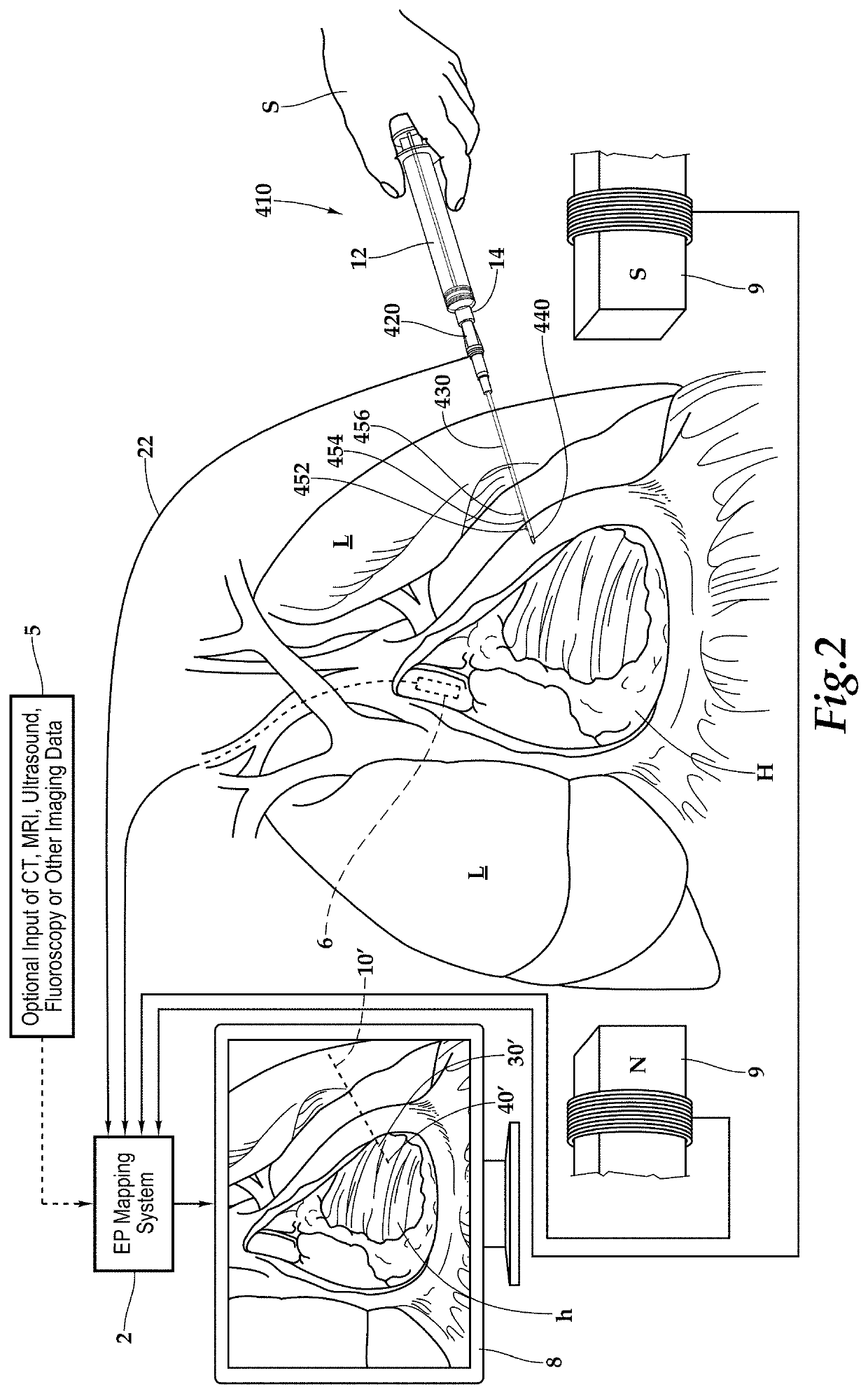
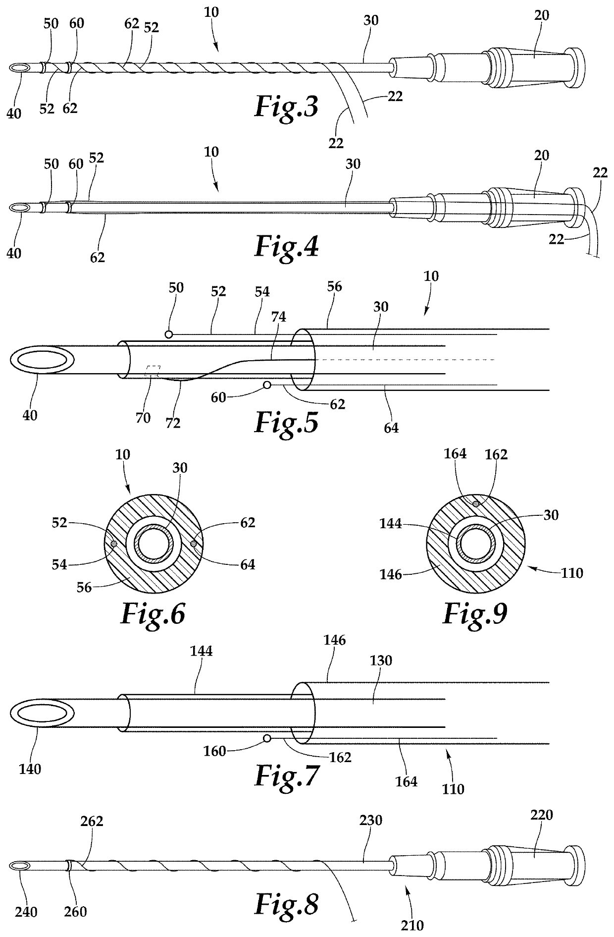
[0113]Referring to the drawings, wherein like reference numerals represent like parts throughout the various drawing figures, reference numeral 1000 is directed to a visualization system for visualization of various subcutaneous spaces (FIGS. 18-20 and 24). The system 1000 utilizes an imaging tool such as an intracardiac ultrasound probe 1020 twice during an intravascular procedure. The probe 1020 or other imaging tool is first used during gaining of vascular access, such as with a percutaneous vascular access needle 10 (FIG. 24), and with the probe 1020 or other imaging tool first covered by an adapter 1030. The probe 1020 or other imaging tool is second used with the adapter 1030 removed by insertion intravascularly and routed to an intra-vascular procedure visualization location. The intracardiac ultrasound probe 1020 is preferably coupled to an EP mapping system 1320 (FIG. 24) both during supra-cutaneous use at the access site and during intravascular use, such as within or adja...


PUM


Abstract

An intracardiac ultrasound catheter or other intravascular imaging tool is provided with an adapter to allow the imaging tool to be used both subcutaneously and supracutaneously, and especially at different stages of a single intravascular procedure. The adapter covers a tip of the imaging tool and preferably includes an echogenic window to facilitate gathering of ultrasound images through this window in the adapter. With the adapter in place, the intracardiac ultrasound can be used supracutaneously, such as at a femoral artery / vein location for imaging to improve vascular access, such as with an appropriate percutaneous needle. The adapter is then removed and the same imaging tool can be inserted percutaneously and intravascularly for use, such as in intracardiac imaging accompanying and intracardiac procedure. The intracardiac ultrasound probe is preferably coupled to an electrophysiology mapping system both when used supracutaneously and when used intravascularly.

Description

CROSS-REFERENCE TO RELATED APPLICATIONS[0001]This application claims benefit under Title 35, United States Code § 119(e) of U.S. Provisional Application No. 62 / 633,793 filed on Feb. 22, 2018 and is a continuation-in-part of U.S. patent application Ser. No. 15 / 713,307 filed on Sep. 22, 2017, which claims benefit under Title 35, United States Code § 119(e) of U.S. Provisional Application No. 62 / 398,394 filed on Sep. 22, 2016 and U.S. Provisional Application No. 62 / 516,556 filed on Jun. 7, 2017.FIELD OF THE INVENTION[0002]This invention relates to use of subcutaneous space visualization tools both for intravascular access, such as with needles, and especially needles fitted with sensors thereon and coupled to electrophysiology mapping systems to identify their position, and imaging tools for intravascular space imaging, and especially intracardiac ultrasonic imaging.BACKGROUND OF THE INVENTION[0003]Cardiac electrophysiology ablation for the treatment of atrial fibrillation has become m...

Claims


Application Information

Patent Timeline
Patent Type & Authority Patents(United States)
IPC IPC(8): A61B34/20A61B8/12A61B8/00A61B8/14A61B18/14A61B8/08A61B18/00A61B90/00A61B17/00
CPCA61B34/20A61B8/0883A61B8/12A61B8/14A61B8/4254A61B8/445A61B8/463A61B18/1477A61B18/1492A61B2017/00053A61B2017/00243A61B2018/00172A61B2018/00178A61B2018/00351A61B2018/00577A61B2018/00839A61B2018/00982A61B2034/2051A61B2034/2053A61B2034/2063A61B2090/3784
Inventor KUSUMOTO, WALTER
Owner KUSUMOTO WALTER
Why Eureka
Try Eureka