Precision optical intracellular near field imaging/spectroscopy technology

a near field imaging and spectroscopy technology, applied in the direction of optical elements, material analysis through optical means, instruments, etc., can solve the problems of cell destruction by electron microscope use, and achieve the optimal impact on biological and medical community, improve the imaging of intact cells, and cost significantly less

Inactive Publication Date: 2006-10-31
NANOPOINTS
View PDF6 Cites 22 Cited by
  • Summary
  • Abstract
  • Description
  • Claims
  • Application Information

AI Technical Summary

Benefits of technology

[0007]The POINT probe collects data from high-resolution imagery, providing a unique imaging tool for the investigation of cells at the sub-cellular and molecular level. These data are incorporated into a signature, which facilitates the molecular analysis of diseases. The current invention has more sensitivity and resolving power than is presently available in microscopy and provides a more detailed understanding of the molecular processes.
[0008]The POINT probe penetrates the cell membrane with a SNOM probe to image the inside of intact cells allowing the POINT probe to collect data from intact cells. This embodiment overcomes the technical difficulties of modifying an existing scanning near-field microscope. The POINT probe greatly improves the imaging of intact cells at the sub-cellular and molecular level without destroying the cell structure.
[0010]The POINT probe characterizes and investigates intact cells with sub-cellular and molecular structures within the context of intact cells. In another embodiment, the POINT probe acts also as an early warning tool facilitating early treatment and eradication of diseased cells.
[0012]The POINT probe allows researchers to gather a richer data set, than that achieved by using conventional methods, from sub-cellular and molecular structures within the context of intact cells. The preferred embodiment improves the imaging of intact cells at the sub-cellular and molecular level without destroying the cell structure. This ability has optimal impact on the biological and medical community to be able to obtain information never before possible.
[0013]One of the major benefits of this system is it costs significantly less than current near-field systems and function with greater capability. The POINT system may be produced as an attachment, so that it can be added to an already existing microscope setup. This means ordinary microscopes, found in most biological and medical labs, to achieve a very high resolution.

Problems solved by technology

Currently confocal microscopes, MRI and ultrasound cannot image to a 50 nm resolution and the use of electron microscopes destroys the cells.

Method used


Image

  • Precision optical intracellular near field imaging/spectroscopy technology
  • Precision optical intracellular near field imaging/spectroscopy technology
  • Precision optical intracellular near field imaging/spectroscopy technology

Examples


Embodiment Construction

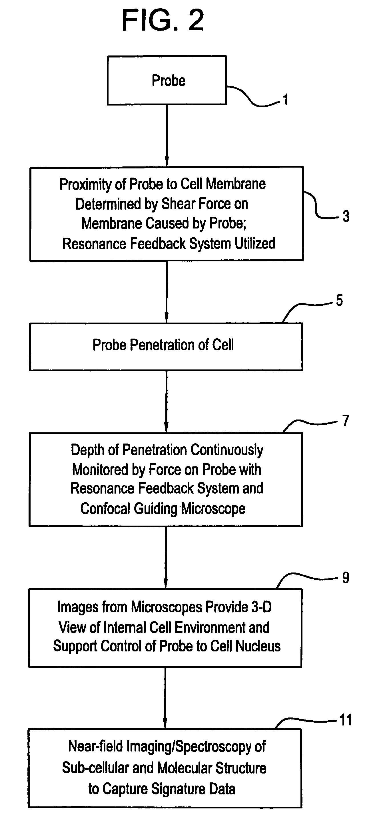
[0025]The preferred embodiment is a method for collecting image data on macro-molecules from intact cells by adapting existing SNOM technology. Although there is some documentation on the use of SNOM on biological samples, the technique has not been perfected. This invention enables intact cells to be penetrated and sub-cellular components to be viewed with an adapted SNOM probe, providing new information about the architecture within a cell and it's molecular composition.

[0026]Most molecular studies are conducted outside of the cell and the ability to view molecular processes within intact cells provides more and better information. The non-invasive probe optimally impacts the field of cell biology, as SNOM technology has not yet been applied to intact, hydrated cells.

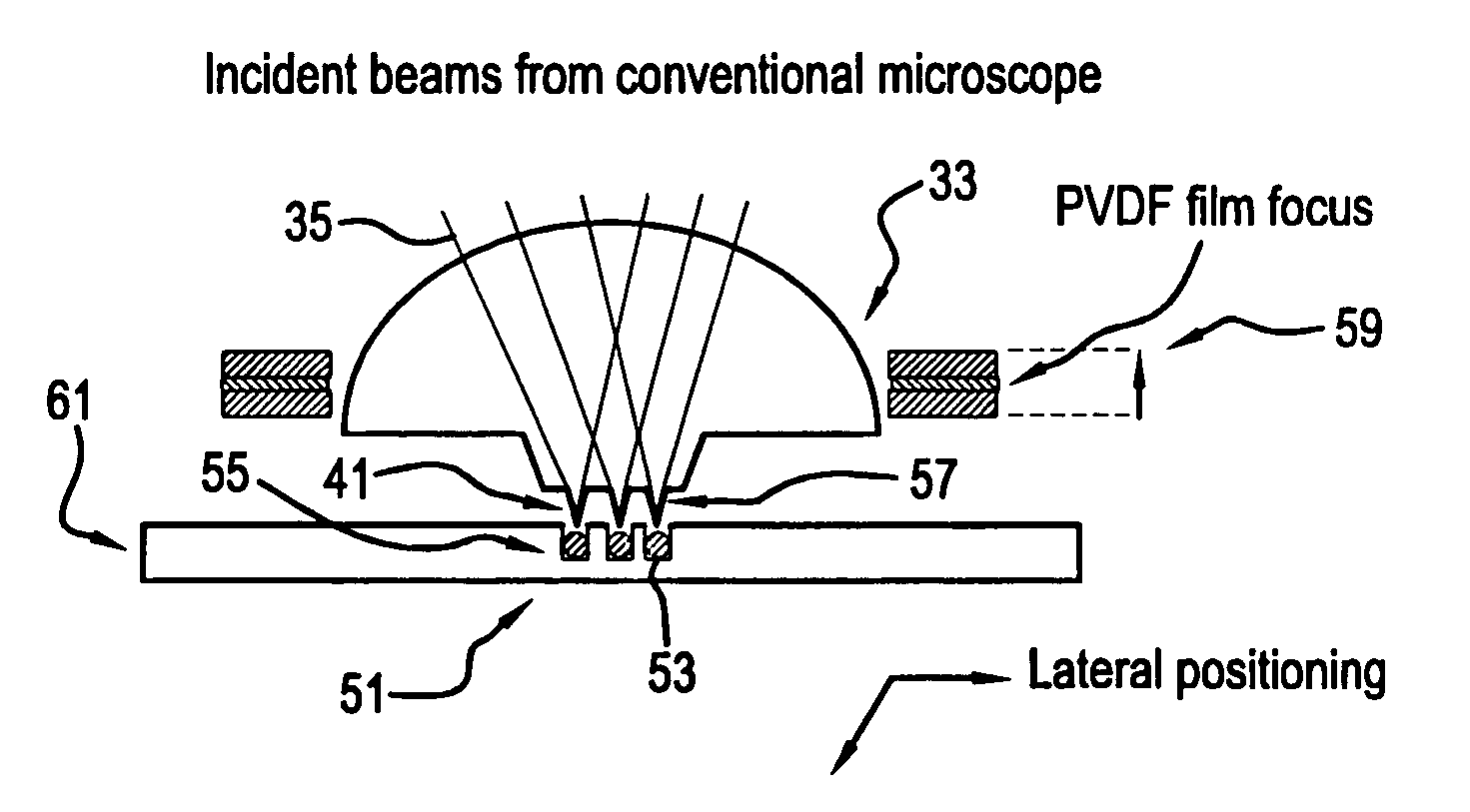
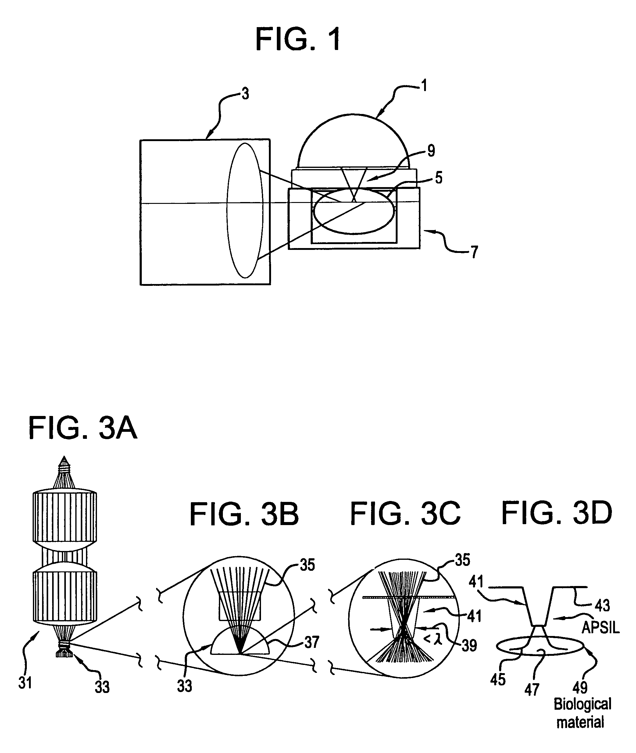
[0027]FIG. 1 shows the basic configuration of POINT with the relative orientation of the modified near-field microscope 1, the POINT probe 9, and the confocal microscope 3 with respect to the cell 5 and the etched cel...


PUM


Abstract

A Precision Optical Intracellular Near Field Imaging / Spectroscopy Technology (POINT / NANOPOINT) is a high-resolution instrument for analyzing and comparing molecular characteristics of cells. A nanosensor array is provided which is capable of imaging inner regions of living cells without destroying its natural environment and providing new information about molecular makeup of cells. The POINT probe collects data from high-resolution imagery, providing an imaging tool for investigating cells at sub-cellular and molecular levels. Data are then incorporated into a signature facilitating molecular analysis of diseases. The POINT probe non-invasively penetrates cell membranes to image insides of intact cells allowing the POINT probe to collect data without destroying cell structures. The probe provides cellular imaging to enable the viewing of both imaging and spectroscopy of internal regions of cells. The POINT system may be attached to existing microscopes to achieve a very high resolution.

Description

[0001]This application claims the benefit of U.S. Provisional Application No. 60 / 331,101, filed Nov. 8, 2001.BACKGROUND OF THE INVENTION[0002]Conventional Scanning Near-Field Optical Microscope (SNOM or NSOM) probes are presently designed to move over the surface of the object being measured, maintaining a constant probe distance from a surface at the nanometer scale while scanning across the surface to obtain nanometer scale resolution images. Scanning near-field microscopy (SNOM) is an optical technique capable of resolving beyond the diffraction limit, where resolutions in the 20 nm range have been demonstrated. The SNOM technology has been applied to cells and to plasma membranes. The high-resolution imagery revealed detail of the cytoskeletal actin not obtainable by any other methods.[0003]SNOM has achieved nano-resolution scales where single molecules have been studied. Near-field techniques circumvent the diffraction limit imposed on conventional light microscopes to obtain s...

Claims


Application Information

Patent Timeline
Patent Type & Authority Patents(United States)
IPC IPC(8): G02B7/04
CPCB82Y20/00B82Y35/00G01Q60/22
Inventor O'CONNELL, DANO'CONNELL-RODWELL, CAITLIN
Owner NANOPOINTS
Why Eureka
Try Eureka