
Method and a MRI apparatus for obtaining images of a target volume of a human and/or animal subject using magnetic resonance imaging (MRI)

a technology of magnetic resonance imaging and target volume, which is applied in the direction of measuring using nmr, magnetic variable regulation, instruments, etc., can solve the problems of human or animal subjects with some medical implants or other non-removable metal inside the body that may not be able to safely undergo an mri examination, and the mri scan typically takes longer

Active Publication Date: 2021-09-07
MAASTRICHT UNIVERSITY +1
View PDF1 Cites 0 Cited by
  • Summary
  • Abstract
  • Description
  • Claims
  • Application Information

AI Technical Summary

Benefits of technology

The invention aims to solve problems in magnetic resonance imaging (MRI) by allowing for faster acquisition of T1-, T2- and diffusion-weighted images. This means that many imaging volumes can be acquired quickly, resulting in quantitative images with less distortion from B1 artifacts. The invention can be used on high and ultra high field scanners without being limited by specific absorption rate. Additionally, the invention uses a technique called stimulated echo acquisition mode (STEAM) to interleave and multiplex acquisitions and increase the number of imaging volumes that can be acquired in the same time sequence, thus reducing filling the dead time and increasing the duty cycle. Overall, the invention improves the speed and accuracy of MRI imaging.

Problems solved by technology

However, compared with CT, MRI scans typically take more time, the clinical environment and the technology used are noisier, and usually require that the subject under examination has to be positioned in a narrow, confined tube (the target area).
In addition, human or animal subjects with some medical implants or other non-removable metal inside the body may be unable to safely undergo an MRI examination.
However, despite its strong advantages, the MRI technique has two disadvantages, which stand in the way of an even greater market share in soft tissue clinical diagnostics: the measurement time and the presence of image artifacts.
First, measurement time for high quality images in MRI is long compared to other commonly used medical imaging techniques.
However, even at high (3T) and ultra-high field strength (UHF, i.e. 7T and beyond) measurement times are long (with ensuing high cost), especially when the increased efficiency is leveraged for more sensitive diagnostics enabled by higher quality scans.
In particular, artifacts due to radiofrequency field (B1) effects, such as transmit and receive field inhomogeneity, distort the clinically important image contrast.
Unfortunately, B1 field inhomogeneity is greater at higher field strengths, which leads to increased B1 induced artifacts at higher fields (3T and 7T).

Method used


Image

  • Method and a MRI apparatus for obtaining images of a target volume of a human and/or animal subject using magnetic resonance imaging (MRI)
  • Method and a MRI apparatus for obtaining images of a target volume of a human and/or animal subject using magnetic resonance imaging (MRI)
  • Method and a MRI apparatus for obtaining images of a target volume of a human and/or animal subject using magnetic resonance imaging (MRI)

Examples


Embodiment Construction

[0010]The invention aims to provide a solution for the above identified problems, allowing an order-of-magnitude speedup in acquiring T1-, T2- and diffusion-weighted images. This high speed of scanning allows many (tens of) imaging volumes to be acquired in short time. These are then suitable for calculating quantitative images which are relatively free of B1 artifacts. As such the invention can be implemented on high and ultra high field (UHF, notably 7 Tesla) scanners without being as limited by specific absorption rate (SAR) as most existing T1-, T2 and diffusion-weighted acquisition techniques are.

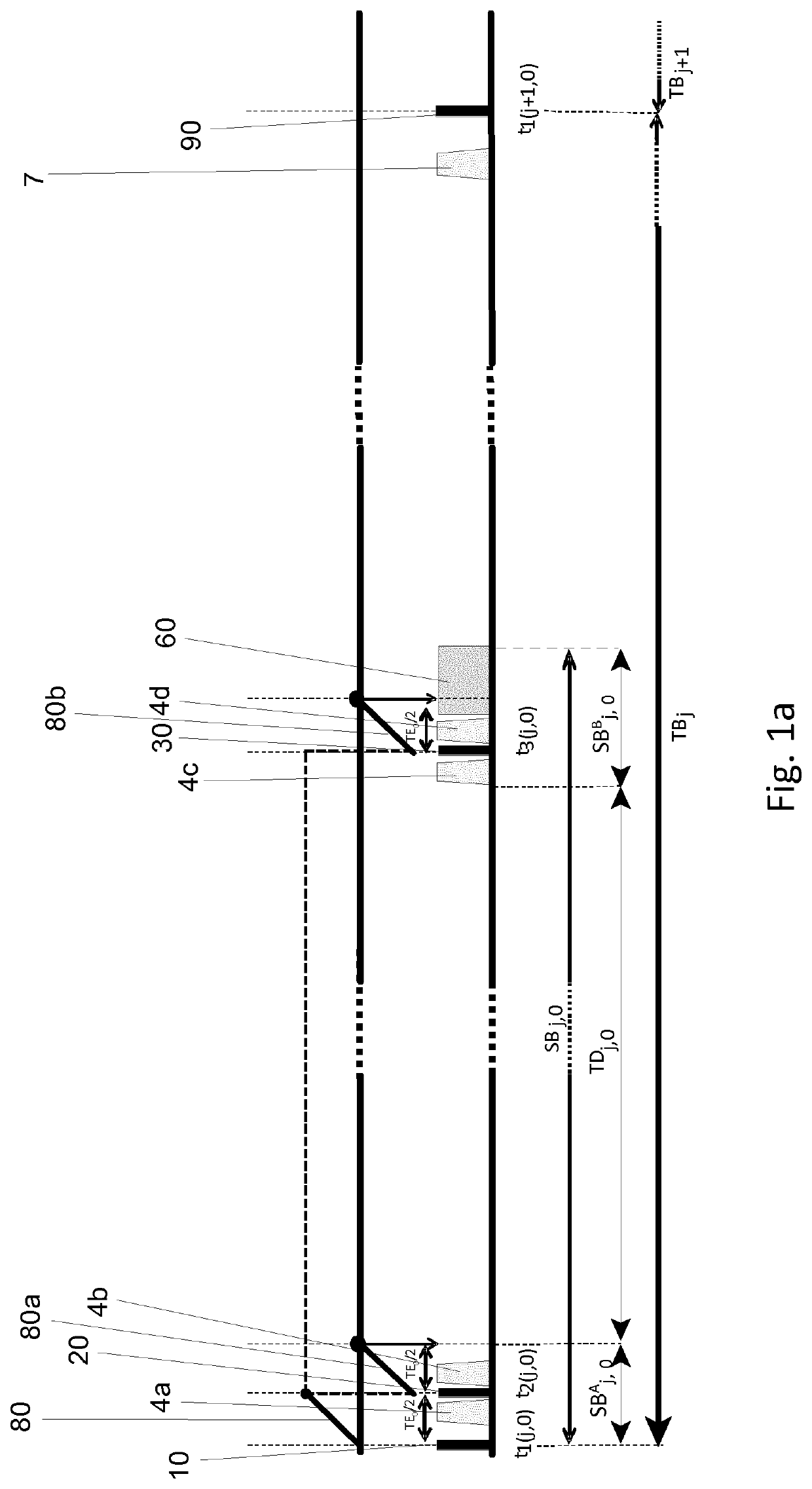
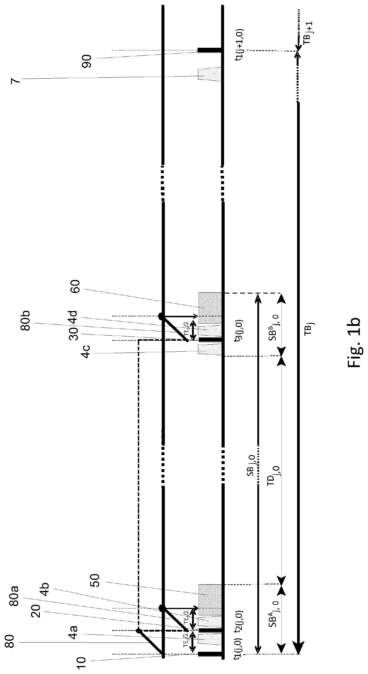
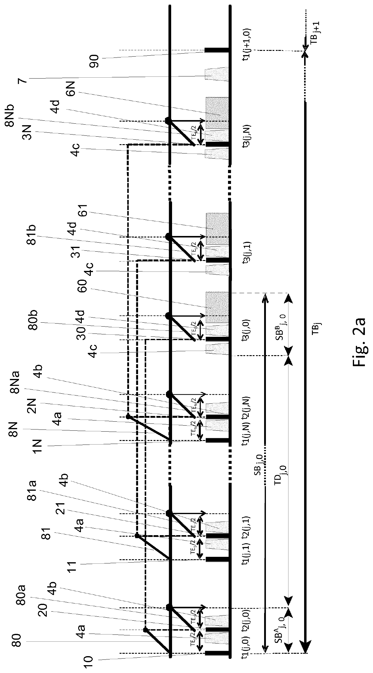
[0011]According to the invention the method for obtaining images of a target volume of a human and / or animal subject using magnetic resonance imaging (MRI) comprising at least the steps of:[0012]A applying, to said human and / or animal subject being positioned in an MRI scanning apparatus, during subsequent total block times TBj with j ϵ [1, 2, . . . , K] a first set of spatially select...


PUM


Abstract

The invention also relates to a MRI apparatus for obtaining images of a target volume of a human and / or animal subject using magnetic resonance imaging (MRI), said MRI apparatus at least comprising: a housing defining a target area for accommodating said human and / or animal subject; at least one main magnet unit and at least one magnetic gradient unit for applying—during use—one or more magnetic field gradients along three independent orthogonal spatial axes in said target area, as well as at least one radiofrequency (RF) pulse generation unit for applying one or more sets of RF pulses towards said target area; a RF receiving unit for acquiring MRI signals possibly having multi-channel spatially sensitive characteristics; and a computer processing unit for processing said acquired MRI signals and for producing said magnetic resonance image data.

Description

CROSS REFERENCE TO RELATED APPLICATIONS[0001]The present application is a 371 U.S. National Phase Entry of pending International Application No. PCT / EP2018 / 077991, filed Oct. 15, 2018, which claims priority to EP 17197326.6, filed Oct. 19, 2017, each of which are herein incorporated by reference in their entireties.FIELD OF THE INVENTION[0002]The invention relates to a method for obtaining images of a target volume of a human and / or animal subject using magnetic resonance imaging (MRI).[0003]The invention also relates to a MRI apparatus for obtaining images of a target volume of a human and / or animal subject using magnetic resonance imaging (MRI), said MRI apparatus at least comprising: a housing defining a target area for accommodating said human and / or animal subject; at least one main magnet unit and at least one gradient magnet unit for applying—during use—one or more magnetic field gradients along three independent orthogonal spatial axes in said target area, as well as at leas...

Claims


Application Information

Patent Timeline
Patent Type & Authority Patents(United States)
IPC IPC(8): G01V3/00G01R33/561G01R33/28G01R33/483G01R33/56G01R33/565
CPCG01R33/561G01R33/288G01R33/4835G01R33/5602G01R33/5659
Inventor ROEBROECK, ALARDPOSER, BENEDIKTLAGOS FRITZ, FRANCISCO
Owner MAASTRICHT UNIVERSITY
Learn More