
Minimally-invasive and activity-dependent control of excitable cells

a technology of excitable cells and activity-dependent control, which is applied in the direction of therapy, animal/human proteins, viruses/bacteriophages, etc., can solve the problems of requiring light delivery and rendering such approaches invasive, and achieve the effect of reducing the extent of or palliation and reducing the time course of symptoms

Active Publication Date: 2022-02-08
CENTRAL MICHIGAN UNIVERSITY +1
View PDF19 Cites 0 Cited by
  • Summary
  • Abstract
  • Description
  • Claims
  • Application Information

AI Technical Summary

Benefits of technology

This method provides a non-invasive, targeted means to modulate excitable cell activity, reducing the need for implanted devices and enabling precise control over cell populations, effectively treating conditions associated with abnormal bursting patterns.

Problems solved by technology

Moreover, delivery of light often requires the use of chronic implanted devices, which may be fully or partially implanted.
If automatic control of optogenetic control is desired in a device, additional elements, such as implanted electrodes, may be required, rendering such approaches invasive.

Method used


Image

  • Minimally-invasive and activity-dependent control of excitable cells
  • Minimally-invasive and activity-dependent control of excitable cells
  • Minimally-invasive and activity-dependent control of excitable cells

Examples


example 1

Expression of a Luminescent Protein

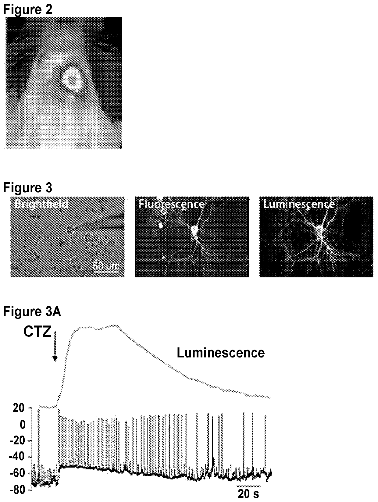
[0176]This example demonstrates a targeted expression of Gaussia luciferase (GLuc) in mouse brain. Several weeks later, coelenterazine was administered intravenously to the mouse, and an image of the mouse's head was taken with IVIS® imaging system (FIG. 2). As shown in FIG. 2, GLuc was expressed in neocortical cells of right hemisphere only.

example 2

rization and Depolarization of Neocortical Neurons

[0177]This examples demonstrates modulation of neocortical neurons in vitro by contacting the neurons with CTZ. The neurons were modified to express fusion proteins including a luminescent protein (enhanced Gaussia luciferase (eGLuc)) and either VChR1 or Mac. Successful expression of the full construct in the neocortical neurons was confirmed by observation of the fluorescence of the reporter protein (upper middle panel titled Fluorescence in FIG. 3). Ability of eGLuc to luminesce was unaffected by the inclusion of light-gated ion channel (VChR1 or Mac), as shown in the upper right panel titled Luminescence in FIG. 3). FIG. 3A (depolarization) shows an increase in spontaneous action potential firing upon contacting CTZ with a cell expressing a fusion protein containing eGLuc-VChR1. FIG. 3B (hyperpolarization) shows a suppression of spontaneous action potential firing upon contacting CTZ with a cell expressing a fusion protein contain...

example 3

g Excitable Cell Activity by Expressing a Luminescent Protein and a Light-Gated Ion Channel in the Same Cell

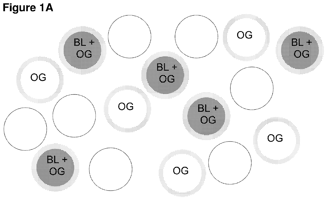
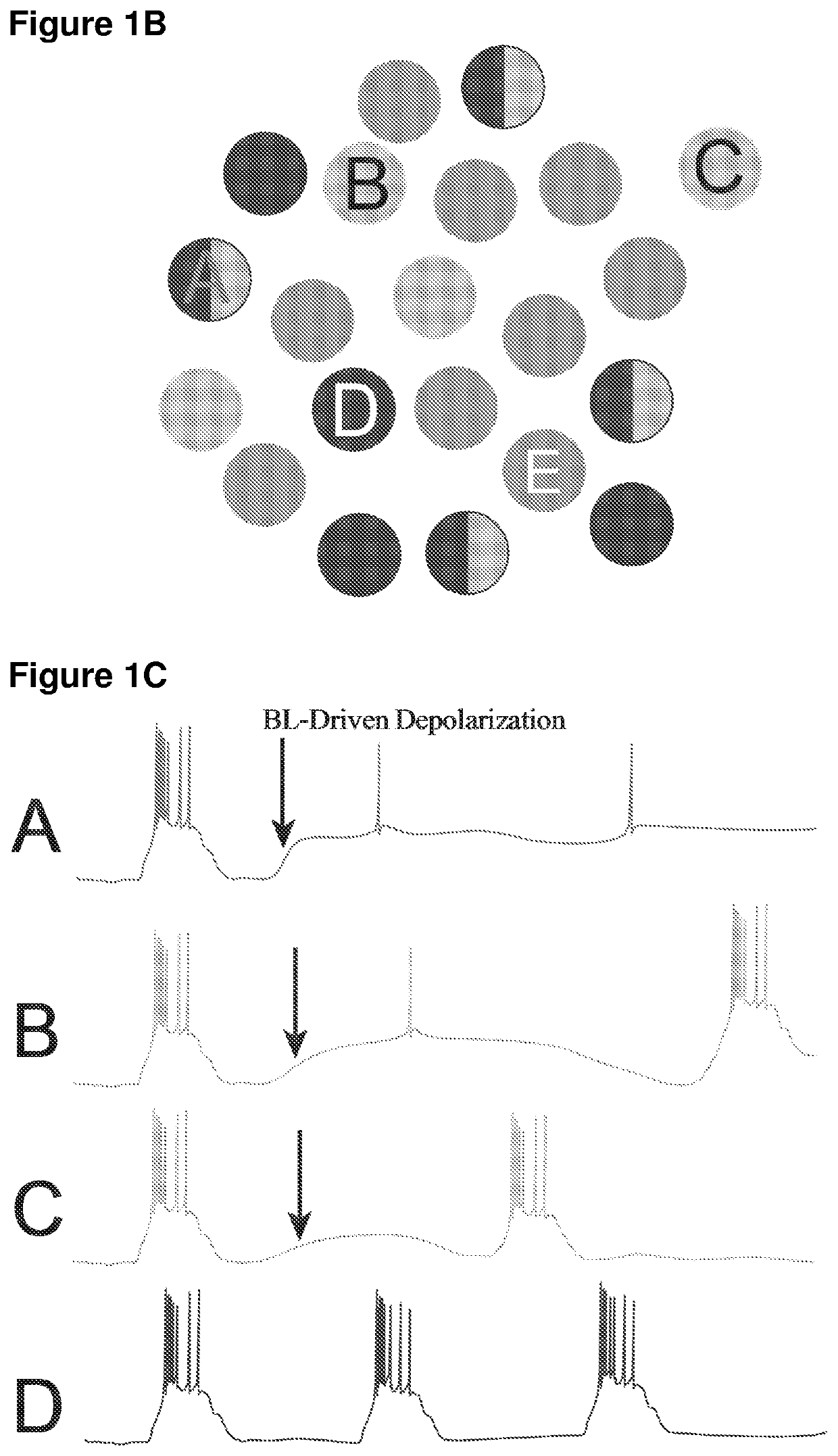
[0178]The use of persistent activity of luminescent protein / light-gated ion channel can be used for burst regulation. This effect can be shown in either single cells, or in cell networks with luminescent proteins expressed in one set of cells and light-gated ion channels in another set of cells. TRN and VPm neurons can be selectively and independently transduced to express a fusion protein containing eGLuc-VChR1 by using methods known in the art. Neurons in brain slices can be visualized in vitro, where high quality current- and voltage-clamp recordings can be made. In particular, the neurons can be neurons in the somatosensory sector of the thalamus, e.g., VPm nucleus and its glutamatergic, excitatory relay neurons, and the adjacent thalamic reticular nucleus and its GABAergic, inhibitory neurons. As shown in FIG. 4A, relay cells receive sensory inputs from lemniscal axons, a...


PUM

PropertyMeasurementUnit
voltagesaaaaaaaaaa
voltagesaaaaaaaaaa
voltagesaaaaaaaaaa

Abstract

The present invention provides a method of bioluminescence-driven optogenetic control of excitable cells. The excitable cell expresses a light-gated ion channel, and a luminescent protein can be expressed either in the excitable cell or in another cell proximal to the excitable cell. The methods of the invention can be used to desynchronize local activity of excitable cells in a mammalian tissue. The methods of the invention can be used to treat a disease or condition in a mammal, the disease or condition being related to bursting. The disease or condition can be Parkinson's disease, epilepsy, a sleep disorder, or a sensory-related disease or condition (e.g., attention deficit disorder or pain). The invention also provides a conjugate of containing a voltage-gated ion channel and a luminescent protein.

Description

CROSS-REFERENCE TO RELATED APPLICATIONS[0001]The present application is a National Stage Entry of PCT / US2016 / 014593 filed Jan. 22, 2016 and claims benefit of U.S. Provisional Patent Application No. 62 / 106,633 filed Jan. 22, 2015, the entire contents of which are incorporated herein by reference.FIELD OF THE INVENTION[0002]The present invention relates to a minimally-invasive modulation of the activity of an excitable cell using bioluminescence-driven optogenetic methods.BACKGROUND[0003]Polarization of excitable cells (e.g., neurons) plays a role in symptoms of various diseases or conditions (e.g., Parkinson's disease, epilepsy, sleep and sensory-related maladies such as pain and attention deficit disorders). While etiologies of these diseases or conditions may differ, their manifestation can involve the same basic processes, such as hyperpolarization of excitable cells. For example, increases in bursting (brief periods of high-frequency action potential activity) are prominent in ep...

Claims


Application Information

Patent Timeline
Patent Type & Authority Patents(United States)
IPC IPC(8): C12N5/0793C07K14/705A61N5/06A01K67/027
CPCC07K14/705A01K67/0275A61N5/062A01K2217/052A01K2217/206A01K2227/105A01K2267/0393C07K2319/60C12N2750/14141
Inventor MOORE, CHRISTOPHER I.HOCHGESCHWENDER, UTELIPSCOMBE, DIANE
Owner CENTRAL MICHIGAN UNIVERSITY
Learn More