Joint hematology and biochemistry point-of-care testing system

a point-of-care testing and joint technology, applied in the field of point-of-care testing (poct) system, can solve the problems of brain damage, blood clot formation, and certain tests that cannot be performed on serum or plasma

Active Publication Date: 2022-05-10
INVIDX CORP
View PDF74 Cites 0 Cited by
  • Summary
  • Abstract
  • Description
  • Claims
  • Application Information

AI Technical Summary

Benefits of technology

[0099]The top portion of the sample storage well of the alternate cartridge (B) as described above may comprise a boss for increasing the sample storage well storage capacity. Furthermore, the cap may be one of a hinged cap and a screw-type cap.
[0116]The system provided herein, comprising an analyzer and a cartridge, provides a compact and versatile POCT system that is effective for use within a variety of health care settings, for example, within space-limited Emergency Department (ED) of hospitals and in the vehicles used by first responders. The system described herein may also be used to determine multiple properties, for example, one or more than one analyte quantity per unit volume of blood and at least one formed element quantity per unit volume of blood, within a single blood sample for example, a capillary blood sample (i.e., a pin-prick blood sample), thereby reducing the need to obtain additional blood samples. By consolidating several different tests in a single, user-friendly POCT analyzer, the system may be operated by non-laboratory personnel.

Problems solved by technology

They also initiate the formation of blood clots.
Due to the complexity of blood, certain tests can only be performed on serum or plasma.
Very high levels of bilirubin in neonates may cause brain damage.
Some developing countries rely on manual blood cell counting, which is time consuming and errorprone.
Although the blood cell counting using the “Coulter Principle” is still very popular in clinical labs, the size of the analyzers limits the use of the “Coulter Principle” for POCT.
POCT has improved patient care in several areas including the Emergency Department (ED) of hospitals, but the ED is usually very busy and may have space for implementing one POCT analyzer, and practical issues (physical constraints) may limit implementation of more than one POCT analyzer.

Method used


Image

  • Joint hematology and biochemistry point-of-care testing system
  • Joint hematology and biochemistry point-of-care testing system
  • Joint hematology and biochemistry point-of-care testing system

Examples


first embodiment

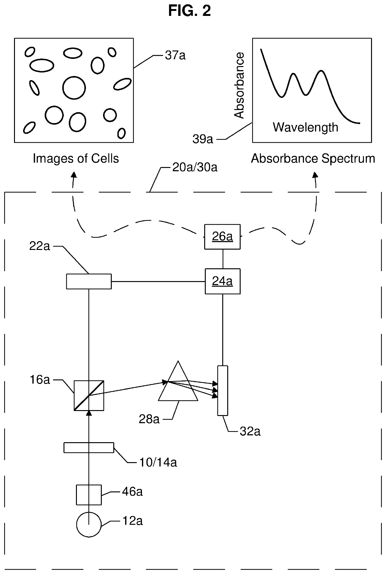
[0212]a system 30a for measuring one or more analyte quantities per unit volume of blood (i.e., the concentration of the analyte) using spectroscopic technique, and one or more formed element quantities per unit volume of blood (i.e., a cell count) using an imaging technique, is illustrated in FIG. 2. System 30a comprises a source of EMR 12a, which may represent a polychromatic source of EMR or a plurality of monochromatic EMR. Depending on the source of EMR, a collimation system 46a may be used to produce substantially parallel rays of EMR for interrogating or illuminating the blood sample in an optical chamber (see 13 in FIGS. 10H & 10K) of a cartridge 10 (10 may be considered a generic cartridge, and could be at least one of 10A, 10B, 10C or 10D, or a similar cartridge described in the prior art) in a receptor 14a of an analyzer 20a (see FIGS. 8A-8C).

[0213]The EMR transmitted through the blood sample in the optical chamber 13 of a cartridge 10 is referred to as emerging EMR. The ...

second embodiment

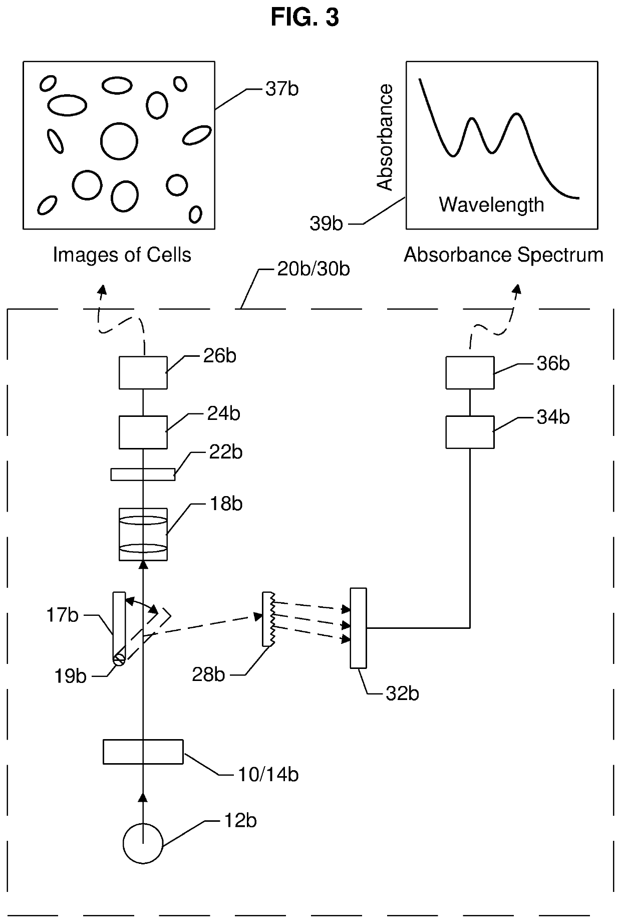
[0236]a system 30b for measuring one or more analyte quantities per unit volume of blood (i.e., the concentration of the analyte) using a spectroscopic technique, and one or more formed element quantities per unit volume of blood (i.e., a cell count) using an imaging technique is illustrated in FIG. 3. The most significant differences when compared with system 30a are:[0237]1) instead of using a beam splitter 16a, a pivotal mirror 17b is used to direct EMR transmitted through the blood sample, to either detector 22b or detector 32b, depending on the position of the mirror;[0238]2) system 30b comprises a magnification system 18b for projecting a real enlarged image on to the detector 22b; and[0239]3) instead of a prism 28a, the EMR dispersing element is a transmission grating 28b.

[0240]The differences between the first set of emerging EMR and the second set of emerging EMR are: a) each set emerges from the sample at a different time, depending on the position of the pivotal mirror 1...

third embodiment

[0241]a system 30c is illustrated in FIG. 4. The most significant differences when compared with system 30b are:[0242]1) a bifurcated optical fiber comprising one or more strands of optical fiber is used as a beam splitter; and[0243]2) instead of a transmission grating 28b, the EMR dispersing element is a reflecting grating 28c.

[0244]The bifurcated optical fiber may be designed so that the magnitude of the first set of emerging EMR and the magnitude of the second set of emerging EMR are optimized to produce accurate measurements of the one or more cell counts and the one or more analyte concentrations.

[0245]A fourth embodiment of a system 30d is illustrated in FIG. 5. The most significance differences when compared to the first three embodiments 30a, 30b and 30c illustrated in FIGS. 2, 3 and 4 respectively are:[0246]1) multiple sources of EMR (12′ and 12d″ are shown as examples, but others may be arranged in a circular manner, with respect to an axis represented by the direction of...


PUM

PropertyMeasurementUnit
wavelengthsaaaaaaaaaa
angleaaaaaaaaaa

Abstract

A joint point-of-care testing (POCT) analyzer, and a system comprising an analyzer and a cartridge, for measuring one or more analyte quantities per unit volume of blood and one or more formed element quantities per unit volume of blood, is described. Examples of formed elements of blood are red blood cells and white blood cells, and cell counts are determined by imaging using a two-dimensional multi-channel detector. Examples of analytes are hemoglobin and bilirubin, and hemoglobin and bilirubin concentrations are determined by spectroscopy using a one-dimensional multi-channel detector. Other examples of analytes are electrolytes, and electrolyte concentrations may be determined using biosensors incorporated in the cartridges.

Description

FIELD OF THE INVENTION[0001]The invention relates to a point-of-care testing (POCT) system that consolidates analytical tests in a single analyzer.BACKGROUND OF THE INVENTION[0002]The result of reaction between a liquid sample and one or more reagent depends on the quantity of the one or more reagent and the volume of liquid sample. Although any type of liquid sample is implied, serum, plasma and blood (also referred to as whole blood) are samples of particular interest. The reagent is preferably in a dry form, in order to avoid dilution of the sample. When blood is allowed to clot and the sample is centrifuged, the yellow liquid that sits on top of the blood clot is called serum. If the blood is collected in a tube containing an anticoagulant, for example heparin, and the blood centrifuged, the cells and cell fragments, referred to as formed elements, are separated from a yellow liquid called plasma, which sits on top of the formed elements. The plasma is usually about 90 percent w...

Claims


Application Information

Patent Timeline
Patent Type & Authority Patents(United States)
IPC IPC(8): G01N33/80G01N33/569G01N33/555G01N33/49
CPCG01N33/80G01N33/49G01N33/555G01N33/56972G01N2496/05G01N2496/15
Inventor SAMSOONDAR, JAMES
Owner INVIDX CORP
Why Eureka
Try Eureka