Non-invasive method for monitoring transplanted organ status in organ-transplant recipients

a transplant recipient and non-invasive technology, applied in the field of medical diagnostics, can solve the problems of high cost of hospitalization, difficult frequent monitoring of transplanted grafts, and significant medical risks

Active Publication Date: 2022-10-25
ACRANNOLIFE GENOMICS PVT LTD
View PDF7 Cites 0 Cited by
  • Summary
  • Abstract
  • Description
  • Claims
  • Application Information

AI Technical Summary

Problems solved by technology

Frequent monitoring of a transplanted graft is difficult as it requires highly invasive procedures like tissue-biopsy, regarded as the “Golden Standard” for determining rejection status of transplant graft and confirmation of graft-rejection, the method is highly invasive necessitating expensive hospitalization, and also carries significant medical risks, including internal bleeding and biopsy-induced transplant graft rejection.
Also, the conventional method for detection of rejection is difficult, expensive, and risky, and do not have adequate specificity and sensitivity to elucidate the rejection status of a transplanted organ.
In addition, this method suffers from many problems including high costs, variability in tissue biopsy between physicians, and severe patient discomfort.
Medical management of transplantation is one of the most challenging and complex specialities in modern medicine.
Not all transplanted organ survives in the new body of a transplant recipient and thus, in a few cases, the organ gets rejected by the transplant-recipient, thereby creating critical, life-threatening medical emergency conditions.
The important challenge for medical management in transplants is the management of graft-rejection that occurs mostly due to immunological response of the recipient's body against the transplanted organ.
However, these methods also pose limitations as they tend to produce high false positive results due to the complex cross-reactivity of various immune responses and are based on tissue-specific gene expression signals.
However, the ddcfDNA is present in minute quantity, whereas the background DNA is present in abundance and poses a limitation to be used as a tool.

Method used


Image

  • Non-invasive method for monitoring transplanted organ status in organ-transplant recipients
  • Non-invasive method for monitoring transplanted organ status in organ-transplant recipients

Examples


example 1

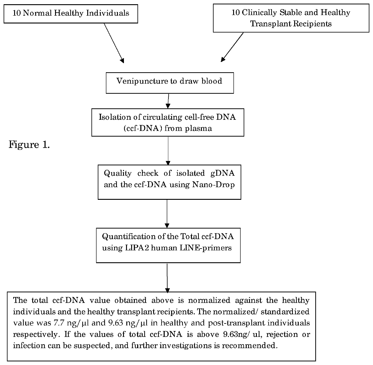
[0037]STEP-1: Sample Collection and Separation of Plasma

[0038]5 ml of peripheral blood was collected by venipuncture into an EDTA-coated collection tube both from the donor and the recipient.

[0039]Urine samples were prepared by allowing it to stand until all the debris is settled and the clear upper layer is taken for analysis.

[0040]From this, 2 ml of blood was centrifuged at 5000 rpm for 10 min and plasma was carefully removed from the top and stored at −20° C. for further use.

[0041]STEP-2: Isolation of Circulating Cell-Free DNA, Genomic DNA:

[0042]2(a) Isolation of Circulating Cell-Free DNA (ccfDNA):

[0043]The circulating cell-free DNA (ccfDNA) is isolated from plasma following the manufacturer's instructions of EpiQuik Circulating cell-free DNA Isolation kit, Epigentek Group Inc. USA. The ccfDNA was isolated by adding 0.5 ml of plasma with 24 μl of ccfDNA capture Enhancer; 900 μl of Capture Buffer and 50 μl ccfDNA Capture Beads into 1.7 ml micro centrifuge tube. The solution was mi...

example 2

[0049]PCR Amplification and Determination of Total ccfDNA Value:

[0050]Total ccfDNA was quantified using multi locus LIPA2 regions. LIPA2 is a human Long Interspersed Element (LINE) of the class L1, that is well interspersed throughout the human genome. Reaction mixture for each LIPA-qPCR (90 bp and 222 bp amplicons) contained 3 μl DNA template, 0.5 μl of the each forward and reverse primer, 0.4 μl Rox as passive reference dye, 10 μl SYBR Green Master Mix (KAPA) and made up to total reaction volume of 20 μl with 95° C. for 1 min, followed by 40 cycles of 95° C. for 15 s. and annealing at 64° C. for 1 min in Stepone plus Real-Time PCR. System (Applied Biosystems, USA).

[0051]LIPA-qPCR reactions were standardized using 10 healthy control samples to yield the total ccfDNA results. The “control-value” is an average of total ccfDNA value of the ccfDNA levels of 10 healthy control and used as the standard. “Control Value” of the total ccfDNA of healthy subject is <7.7 ng / μl. The total ccfDN...

example 3

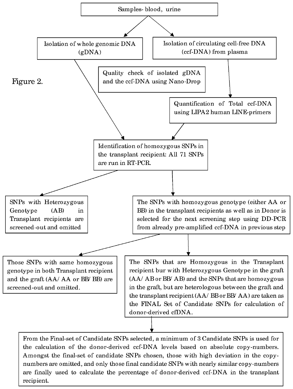
[0053]PCR Amplification and Determination of the Donor-Derived ccfDNA.

[0054]The donor derived ccfDNA amplification is performed by DDPCR for the identification of the organ transplant status in the transplant recipient. Firstly, screening of all the High-MAF SNPs was done in both genomic DNA as well as the circulating cell-free DNA using Real-Time PCR. In this step, the SNPs that have heterozygous genotype in the recipient are filtered out and eliminated, since they cannot be used for quantification in DDPCR. In the next step, the pre-amplified ccfDNA is used as the template, and the candidate SNPs are filtered out and the assay is set for the individual patient. The SNPs which are homozygous in the recipient and that could be either heterozygous or homozygous on the allele in the graft and is preferably heterologous between the recipient and the graft is chosen as the final candidate SNPs. This step is performed as a DDPCR assay.

[0055]All homozygous SNPs were subsequently used to g...


PUM

PropertyMeasurementUnit
volumeaaaaaaaaaa
volumeaaaaaaaaaa
volumeaaaaaaaaaa

Abstract

The present invention pertains to a non-invasive method for monitoring transplanted organ status in organ-transplant recipients by determining the ratio of donor derived marker sequences to the marker sequences of the transplant recipient from circulating cell free DNA of the transplant recipients using digital droplet PCR. The invention also determined a normalized threshold value of the total circulating cell free nucleic acids healthy as well as in post-transplantation patients for assessing and monitoring transplanted organ status in organ-transplant recipients.

Description

SEQUENCE LISTING[0001]This application contains a sequence listing filed in electronic form as an ASCII.txt file entitled “KNS-002-US-Seq-listing-txt.txt” created on Dec. 14, 2018; and the size is 28 KB. The content of the sequence listing is incorporated herein in its entirety.FIELD OF INVENTION[0002]The invention pertains to the field of medical diagnostics with special reference to non-invasive method for monitoring transplanted organ status in organ-transplant recipients.BACKGROUND[0003]Transplantation is a life-extending medical procedure and the most preferred treatment option for end-stage organ damages as long-term mortality is almost 68-70% lower in patients receiving an organ-transplant than those patients who did not receive a transplant. Frequent monitoring of a transplanted graft is difficult as it requires highly invasive procedures like tissue-biopsy, regarded as the “Golden Standard” for determining rejection status of transplant graft and confirmation of graft-rejec...

Claims


Application Information

Patent Timeline
Patent Type & Authority Patents(United States)
IPC IPC(8): C12Q1/6883
CPCC12Q1/6883C12Q2600/112C12Q2600/156
Inventor RAMANI, AVINASH
Owner ACRANNOLIFE GENOMICS PVT LTD
Why Eureka
Try Eureka