Process for discriminating and counting erythroblasts

a technology of erythroblasts and counting methods, applied in chemical methods analysis, dead animal preservation, instruments, etc., can solve the problems of inability to accurately determine erythroblasts, and easy damage to cell membranes of leukocytes as well as erythroblasts

Inactive Publication Date: 2002-01-17
SYSMEX CORP
View PDF0 Cites 24 Cited by
  • Summary
  • Abstract
  • Description
  • Claims
  • Application Information

AI Technical Summary

Problems solved by technology

However, such a method requires not only time-consuming, complicated pretreatment of blood for examination but also considerable expertise and skills for obtaining accurate results.
In these methods, however, cell membranes of leukocytes as well as erythroblasts become easily damaged especially in a hematologic sample with increasing time after collection of blood.
As a result, erythroblasts cannot be accurately determined.
Especially, when lymphoid cells are damaged, it becomes more difficult to distinguish erythroblasts from damaged lymphoid cells clearly and thus the presence of erythroblasts cannot be detected accurately.
Besides, it is difficult to accurately discriminate and count erythroblasts in a sample containing lymphoblasts or a sample in which chemotherapy or the like has made membranes of leukocytic cells ready to be damaged by an hemolytic agent, even if the sample does not go through change with time.
This method uses two antibodies and one fluorescent dye as reagents and has a disadvantage in that the reagents are expensive.
Since the method can examine immature, nucleated red blood cells only with the combination of these specific antibodies with the specific dye, it is impossible to analyze erythroblasts cheaply by use of this method.
However, since this publication lacks description about the distinction of erythroblasts from platelets and debris, it is difficult to count erythroblasts exactly by this method.
Thus absolute counting is difficult.
Moreover, since this centrifugal cleaning involves complicated operation, results of detection vary significantly depending on the skill of an examiner.
However, such method has not been established so far.
If the pH is too low, the reagent fluid does excessive damage to not only erythrocytes but also leukocytes, erythroblasts and the fluorescent labeled antibody specific to leukocytes.

Method used


Image

  • Process for discriminating and counting erythroblasts
  • Process for discriminating and counting erythroblasts
  • Process for discriminating and counting erythroblasts

Examples


example 1

[0069] First, reagents having the following compositions were prepared.

[0070] Fluorescent labeled antibody:

[0071] FITC labeled anti-CD45 antibody

[0072] First reagent fluid (pH 3.0, osmotic pressure 16 mOsm / kg.multidot.H.sub.2O):

[0073] Buffering agent

[0074] Citric acid monohydrate, 2.10 g / l

[0075] Disodium hydrogenphosphate, 0.56 g / l

[0076] Nucleotide fluorescent dye

[0077] Propidium iodide, 100 mg / l

[0078] Purified water

[0079] Second reagent fluid (pH 7.5, osmotic pressure 420 mOsm / kg.multidot.H.sub.2O):

[0080] Buffering agent

[0081] Sodium dihydrogenphosphate dihydrate, 0.95 g / l

[0082] Disodium hydrogenphosphate, 6.24 g / l

[0083] Osmolarity compensating agent

[0084] Sodium chloride, 10.2 g / l

[0085] Purified water

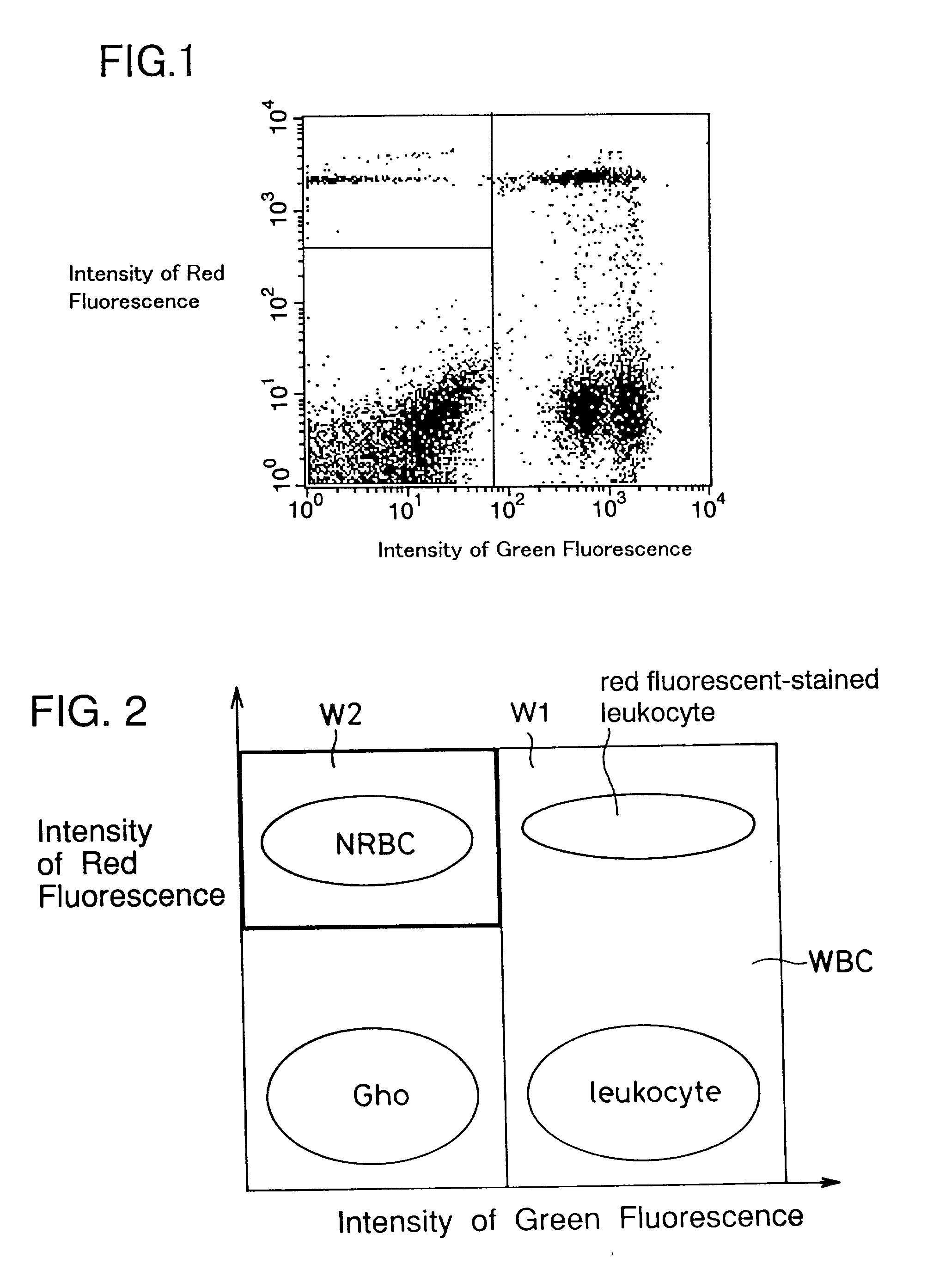
[0086] Fifty (50) .mu.l of blood from a patient was added to peripheral blood to produce a hematologic sample. These bloods had been treated with an anticoagulant. Ten (10) .mu.l of the above FITC labeled anti-CD45 antibody were added to the hematologic sample. This mixture was incuba...

example 2

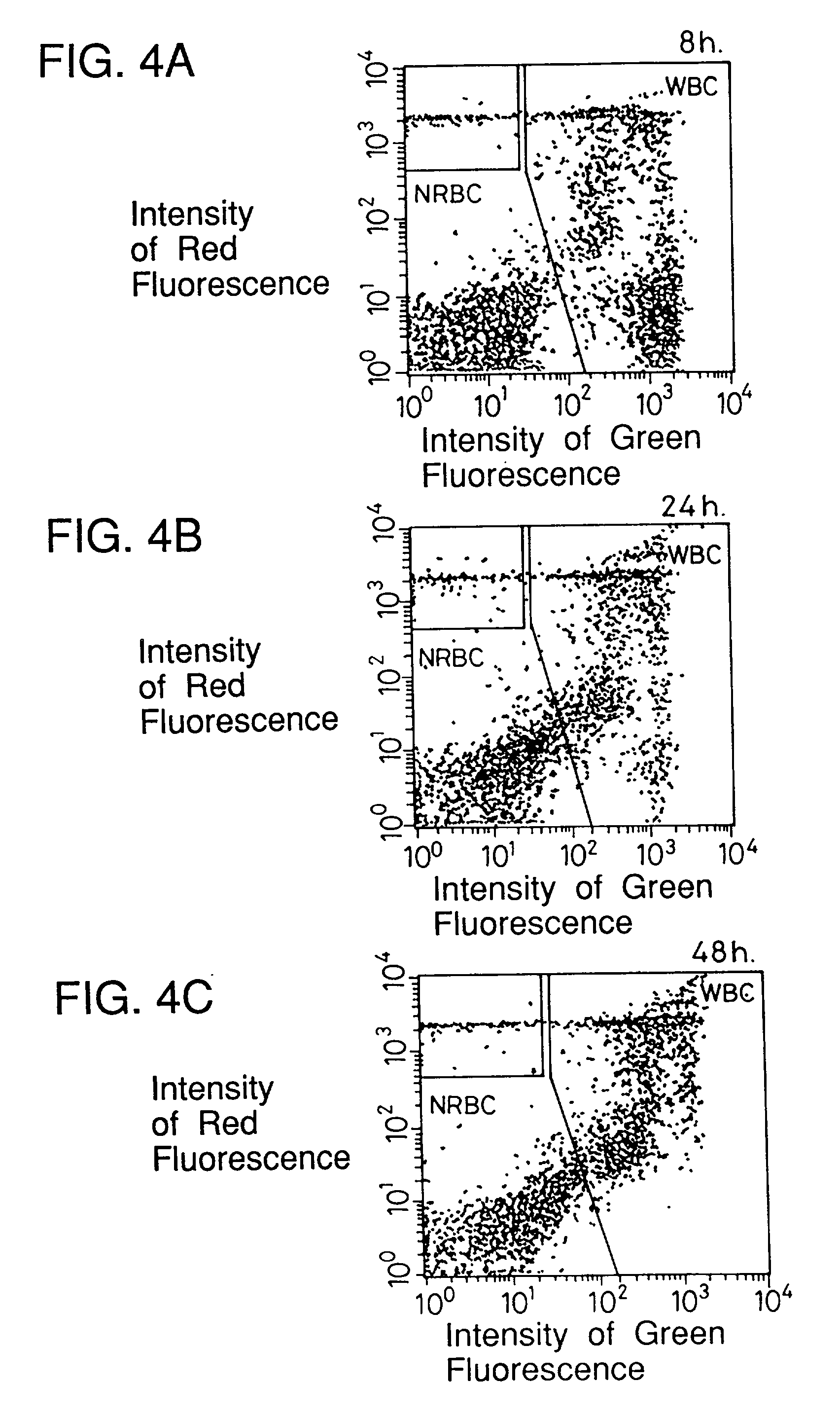
[0094] Blood containing erythroblasts from two patients other than the patient in Example 1 and peripheral blood (preserved at room temperature for 8 hours, 24 hours, 48 hours after collection) were examined about leukocytes and erythroblasts in the same manner as in Example 1. The results are shown in FIGS. 4A to 4C and FIGS. 5A to 5C.

[0095] The ratio of the erythroblasts to the leukocytes was also calculated for each of the samples. The results are shown in Table 1.

1 TABLE 1 after 8 hours after 24 hours after 48 hours Sample 1 3.6% 3.6% 3.6% Sample 2 6.1% 6.1% 6.7%

[0096] FIGS. 4A to 4C and 5A to 5C and Table 1 show that the passing of time hardly affects the results of examination according to the present invention.

example 3

[0097] Reagents having the following compositions were prepared.

[0098] Fluorescent labeled antibody:

[0099] FITC labeled anti-CD45 antibody

[0100] First reagent fluid (pH 3.0, osmotic pressure 16 mOsm / kg):

[0101] Buffering agent

[0102] Citric acid monohydrate, 2.10 g / l

[0103] Disodium hydrogenphosphate, 0.56 g / l

[0104] Nucleotide fluorescent dye

[0105] Propidium iodide, 1 mg / l

[0106] Purified water

[0107] Second reagent fluid (pH 7.5, osmotic pressure 420 mOsm / kg):

[0108] Buffering agent

[0109] Sodium dihydrogenphosphate dihydrate, 0.95 g / l

[0110] Disodium hydrogenphosphate, 6.24 g / l

[0111] Osmolarity compensating agent

[0112] Sodium chloride, 10.2 g / l

[0113] Purified water

[0114] Fifty (50) .mu.l of blood from a patient was mixed with ten (10) .mu.l of the above FITC labeled anti-CD45 antibody. These bloods had been treated with an anticoagulant. This mixture was incubated at room temperature for about 15 minutes.

[0115] Then, 500 .mu.l of the first reagent fluid were added to the mixture, which wa...


PUM

PropertyMeasurementUnit
concentrationaaaaaaaaaa

Abstract

A method for discriminating and counting erythroblasts comprises the steps of: (i) staining leukocytes in a hematologic sample by adding a fluorescent labeled antibody capable of binding specifically with leukocytes to the hematologic sample; (ii) raising the permeability only of cell membranes of erythroblasts in the hematologic sample to a nucleotide fluorescent dye which does not permeate a cell membrane usually, the nucleotide fluorescent dye having a fluorescent spectrum capable of being distinguished from that of a fluorescent labeling compound of the fluorescent labeled antibody in step (i); (iii) staining nuclei of the erythroblasts in the hematologic sample with the nucleotide fluorescent dye; (iv) subjecting the hematologic sample to flowcytometry to detect at least two fluorescent signals from each cell; and (v) discriminating and counting the erythroblasts from difference in intensity between the at least two fluorescent signals.

Description

[0001] 1. Field of the Invention[0002] The present invention relates to a method for discriminating and counting erythroblasts. In particular, the present invention relates to a method for accurately discriminating and counting erythroblasts by flowcytometry.[0003] 2. Description of the Prior Art[0004] The discrimination and counting of erythroblasts are of great use in a field of clinical examination since it provides very useful information for the diagnosis of diseases and prognosis of diseases.[0005] Erythroblasts, which are also called nucleated red blood cells, are normally contained in the bone marrow, but not in peripheral blood except newborns. The appearance of erythroblasts in peripheral blood indicates a possible presence of a disease, such as leukemias, hemolytic anemia, iron deficiency anemia or pernicious anemia, and other non-hematologic / oncolog-ic disorders. Thus the discrimination and counting of erythroblasts are very effective for the diagnosis of any such diseas...

Claims


Application Information

Patent Timeline
Patent Type & Authority Applications(United States)
IPC IPC(8): G01N33/50G01N33/569G01N33/80
CPCG01N33/5094G01N33/56972G01N33/80G01N2015/1006G01N2015/1402Y10T436/107497Y10T436/25125Y10T436/2525Y10T436/25375Y10T436/13Y10T436/108331G01N15/1459
Inventor HOUWEN, BERENDWANG, FU-SHENGTSUJI, TOMOHIROSAKATA, TAKASHIHAMAGUCHI, YUKIO
Owner SYSMEX CORP
Why Eureka
Try Eureka