Analysis of biological samples utilizing a coated solid phase

a solid phase and analysis technology, applied in biological material analysis, instruments, measurement devices, etc., can solve the problems of affecting the deterioration of platelets during storage, affecting the diagnostic field of platelets, and prone to artifactual activation of platelets

Inactive Publication Date: 2002-06-20
HENRY MICHAEL R +2
View PDF2 Cites 11 Cited by
  • Summary
  • Abstract
  • Description
  • Claims
  • Application Information

AI Technical Summary

Problems solved by technology

However, platelets are very prone to artifactual activation when they are centrifuged and handled (Metcalfe, P., Williamson, L. M., Reutelingsperger, C. P., Swann, I., Ouwechand, W. H. & Goodall, A. H. (1997) "Activation during preparation of therapeutic platelets affects deterioration during storage: a comparative flow cytometric study of different production methods."
Artifactual activation has hampered the platelet diagnostic field for decades as th

Method used


Image

  • Analysis of biological samples utilizing a coated solid phase
  • Analysis of biological samples utilizing a coated solid phase
  • Analysis of biological samples utilizing a coated solid phase

Examples


example 2

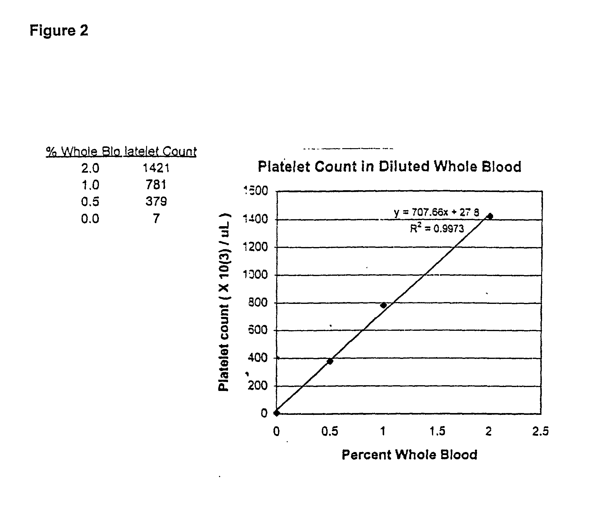
Platelet Capture in Diluted Whole Blood

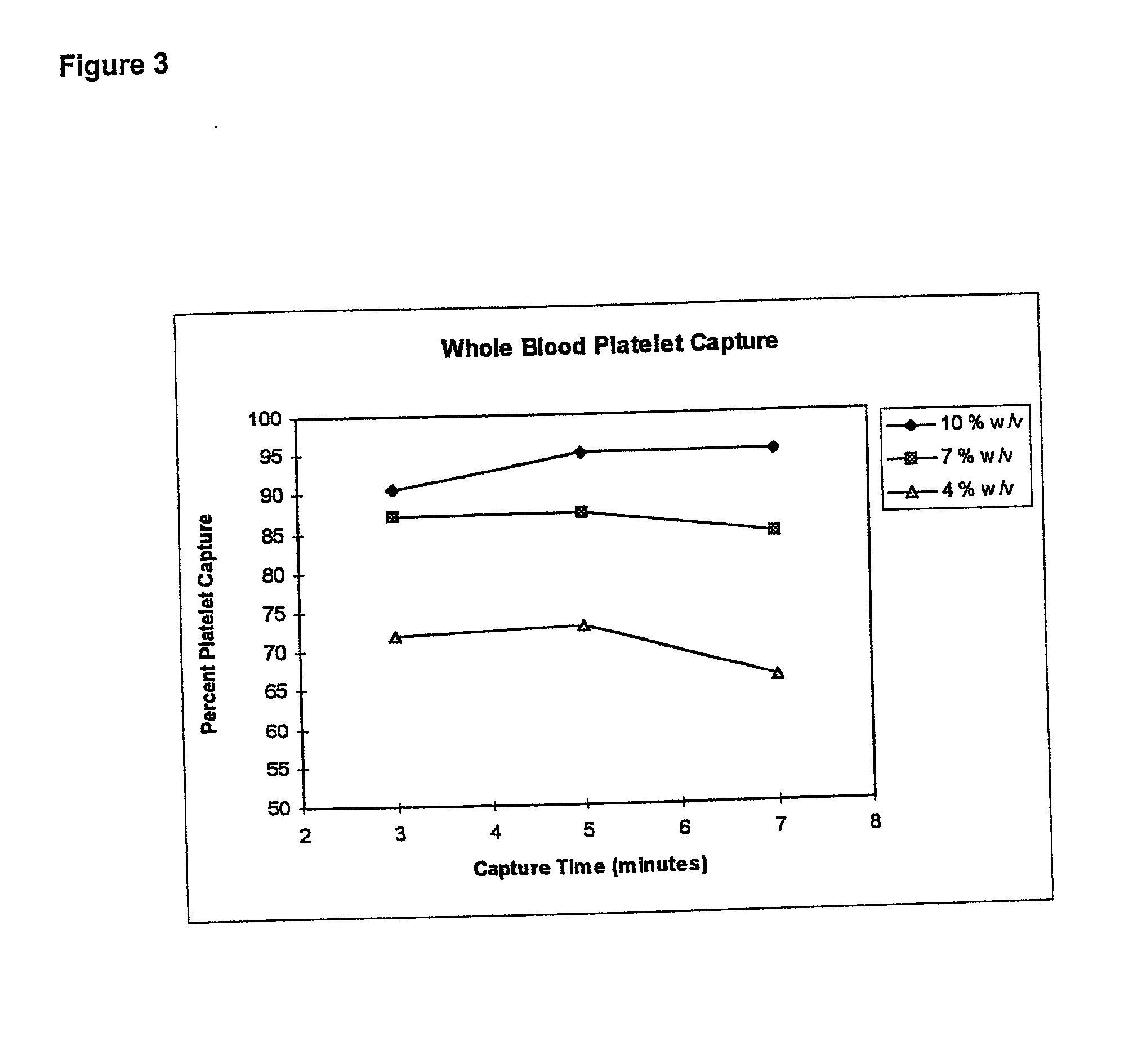
[0066] This example illustrates the dose response of time and solid phase percentage (surface area) to percent platelet capture from whole blood samples.

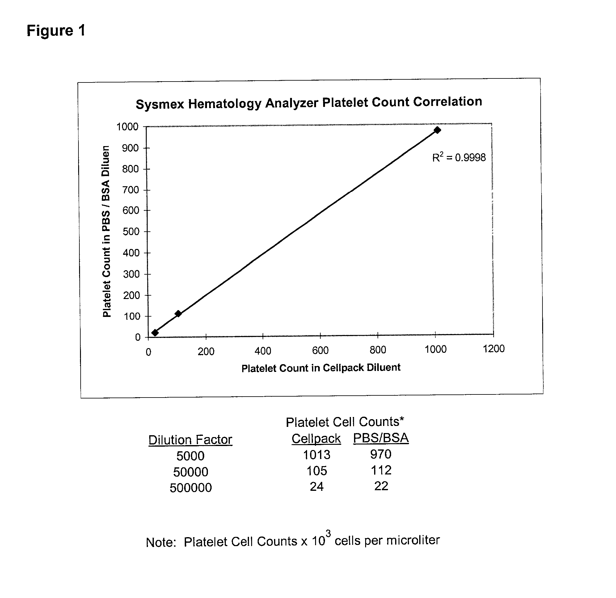
[0067] A multivariant designed experiment was used to examine platelet capture in diluted whole blood. Experimental parameters were: incubation time was about 3-7 minutes; antibody coating concentration was about 6-50 pg; particle concentration was about 4-10% (w / v). The assay was carried out as follows: whole blood was diluted to 2% with phosphate buffered saline (PBS) at pH 7.2 supplemented with 1% bovine serum albumin (BSA). 100 .mu.L of paramagnetic anti-GP1b-coated particles were added to 100 .mu.L of diluted whole blood at room temperature. Following an incubation period, the particles were separated magnetically and the supernatant was removed for testing in a Sysmex F-800 Hematology Analyzer Microcellcounter as previously described in example 1. The platelet concentration per .mu.L was ...

example 3

[0068] This example further illustrates the capture and removal of platelets from whole blood or diluted whole blood.

[0069] Whole blood was obtained from healthy volunteers and collected into centrifuge tubes containing D-phe-pro-arg-chloromethylketone (PPACK, dihydrochloride) anticoagulant. A volume of whole blood was diluted to 2.0% in phosphate buffered saline (PBS), pH 7.4, supplemented with 1% bovine serum albumin (BSA)

[0070] 100 uL of Murine monoclonal antibody anti-GP1b coated paramagnetic microparticles were added to 100 .mu.L of a 2.0% whole blood dilution and incubated at room temperature for 5 minutes. The paramagnetic particles were separated magnetically and the supernatant removed for platelet cell count analysis as previously described in Example 1 with a Sysmex Microcellcounter F-800 Hematology Analyzer.

2 TABLE 2 Avg. - Avg.* bkgd.* No Particle Control 601, 605 601 599 No Particle Control 594, 605 Positive Control 1** 67, 68 59 57 Positive Control 2** 52, 55 Positive...

example 4

Soluble P-selectin Assay in Plasma

[0071] This example illustrates the feasibility of assaying substantially platelet free samples for markers which can be influenced by the presence of physiologically activated platelets within the sample. In this example soluble P-selectin concentrations are measured in substantially platelet free samples to allow for discrimination from membrane P-selectin.

[0072] Recombinant human P-selectin was added to 2.0% platelet-free human plasma sample at the following concentrations: 400, 200, 100, 50, 25, 10 and 1.0 ng / mL. 50 .mu.L of 0.125% (w / v) paramagnetic microparticles (1.5 micron (.mu.m)) coated with an anti-P-selectin murine monoclonal antibody (Mab) (anti-CD62P) were added to 100 .mu.L of each of the aforementioned sample concentrations of recombinant P-selectin in plasma at room temperature and incubated for 10 minutes The microparticles were separated magnetically and the supernatant was removed. The microparticles were washed by re-suspension ...


PUM


Abstract

The invention disclosed herein comprises methods for analyzing a biological sample, such as undiluted or diluted whole blood, as well as fractions thereof, for the presence or absence and/or the concentration of disease-specific and/or other medical condition-specific markers. Such markers may include platelet activation and coagulation activation markers. The methods may comprise combining the biological sample with a coated solid phase and analyzing for the presence or absence and/or the concentration of the markers. The analysis may be performed either before or after separation of the solid phase from the biological sample. The analysis may be performed on the combined components or on any of the separated components. A preferred solid phase may be paramagnetic microparticles coated with antibodies or proteins specific for platelet activation and/or coagulation activation markers.

Description

[0001] This application claims priority from U.S. Provisional Patent Application No. 60 / 222,204, filed Aug. 1, 2000 (Atty. Docket No. MBHB00-555) and U.S. Provisional Patent Application No. 60 / 299,129, filed Jun. 18, 2001 (Atty. Docket No. MBHB00-555-A). All patents, patent applications (published or unpublished) and other scientific or technical writings referred to herein are hereby incorporated by reference to the extent that they are not contradictory.BACKGOUND OF THE INVENTION[0002] 1. Field of the Invention[0003] The invention is related to the field of blood analysis. In particular, blood analysis using a solid phase coated with marker-specific compounds. More in particular, blood analysis wherein the solid phase comprises paramagnetic particles and the marker-specific compounds comprise antibodies, receptors, ligands, proteins, peptides, cytokines, chemokines, small molecules and the like. Even more in particular, the invention is related to the analysis of activated and una...

Claims


Application Information

Patent Timeline
IPC IPC(8): G01N33/543G01N33/566G01N33/86
CPCG01N33/54326G01N2333/705G01N33/86G01N33/566
Inventor HENRY, MICHAEL R.LEAHY, DAVID C.SHEBUSKI, RONALD J.
Owner HENRY MICHAEL R
Why Eureka
Try Eureka