Methods and kits for detecting protein kinases

a protein kinase and kit technology, applied in the field of protein kinase activity detection, can solve the problems of forming tumours, reducing requiring a lot of labor, so as to reduce the amount of atp, reduce the rate of signal decay, and reduce the activity of kinas

Inactive Publication Date: 2002-11-21
LONZA GRP
View PDF0 Cites 14 Cited by
  • Summary
  • Abstract
  • Description
  • Claims
  • Application Information

AI Technical Summary

Benefits of technology

0108] To determine whether there was a concentration dependent effect of kinase activity on reduction in ATP, MAPK-1 was used at concentrations ranging from 1.56 ng/10 .mu.l to 100 ng/10 .mu.l, with serial doubling dilutions from the highest concentrations. The concentration of myelin basic protein used was the same as in the previous experiments. The assays were performed as in the previous two experiments, i.e. using UBI reagents, but with the addition of 110 .mu.l of 200 mM Hepes buffer prior to determination of ATP readings. The results showed a concentration dependent reduction in ATP levels detected with increasing concentrations of the MAPK-1. There was no effect of the enzyme when 1.56 ng was added to each well, however, there was a significant effect at concentrations of 3.13 ng and above (see FIG. 4).
0109] The luciferase enzyme itself is an ATPase that converts ATP to AMP and inorganic phosphate. After the initial increase in light output as a result of the luciferin-luciferase reaction, the light signal begins to decay over time. We examined the light decay with increasing concentrations of ATP up to the 200 .mu.M used in the above experiments. The ATP standards (Sigma, UK) were diluted at serial doubling dilutions from 200 .mu.M down to 3.125 .mu.M per 10 .mu.l added to each well. The standards were diluted in three different buffers, assay dilution buffer from UBI, Tris acetate buffer (pH 7.75) and 200 mM Hepes pH 7.7. 10 .mu.l of the standards, plus a no ATP buffer control, were added in to triplicate wells of the opaque-white 96 well microtitre plates. The experiments were run with duplicate plates, one where 140 .mu.l of the Tris-acetate buffer was added to all wells, and the other where the same volume of 200 mM Hepes buffer. Immediately after the addition of these reagents 20 .mu.l of ATP monitoring reagent was added to all wells. The plate was then placed in a Berthold (RTM) Detection Systems MPL2 luminometer, and the program was set to take 1 second integral readings for each well every 2 minutes. The FIG. 5 graph shows the initial light output (at time 0) and the decay in the light signal observed with the different buffer conditions and the 200 .mu.M stock ATP (final concentration in the well of 12 .mu.M).
0110] Experiments were also carried out to investigate the effect of performing the kinase assay in the presence of luciferin-luciferase reagent (ADR), and its effect upon the rate of signal decay. The enzyme used was JNK-2 (Upstate Biotechnology Inc, USA) at 1 .mu.g per 10 .mu.l added to each well, the c-jun (1-169)-GST substrate (Upstate Biotechnology Inc, USA) was also added at the same concentration. Into each well was added 10 .mu.l of 200 .mu.M ATP standard and 10 .mu.l of Hepes buffer (200 mM). Into control wells the 10 .mu.l of enzyme was replaced by 10 .mu.l of Hepes buffer,

Problems solved by technology

A lack of controlled activity of kinases in cells is believed to lead to the formation of tumours.
As in the case of many of the methods that have been proposed as an alternative to the radioisotope assays, however, the above western

Method used


Image

  • Methods and kits for detecting protein kinases
  • Methods and kits for detecting protein kinases
  • Methods and kits for detecting protein kinases

Examples


example 1--

Determination of the Activity of JNK-1

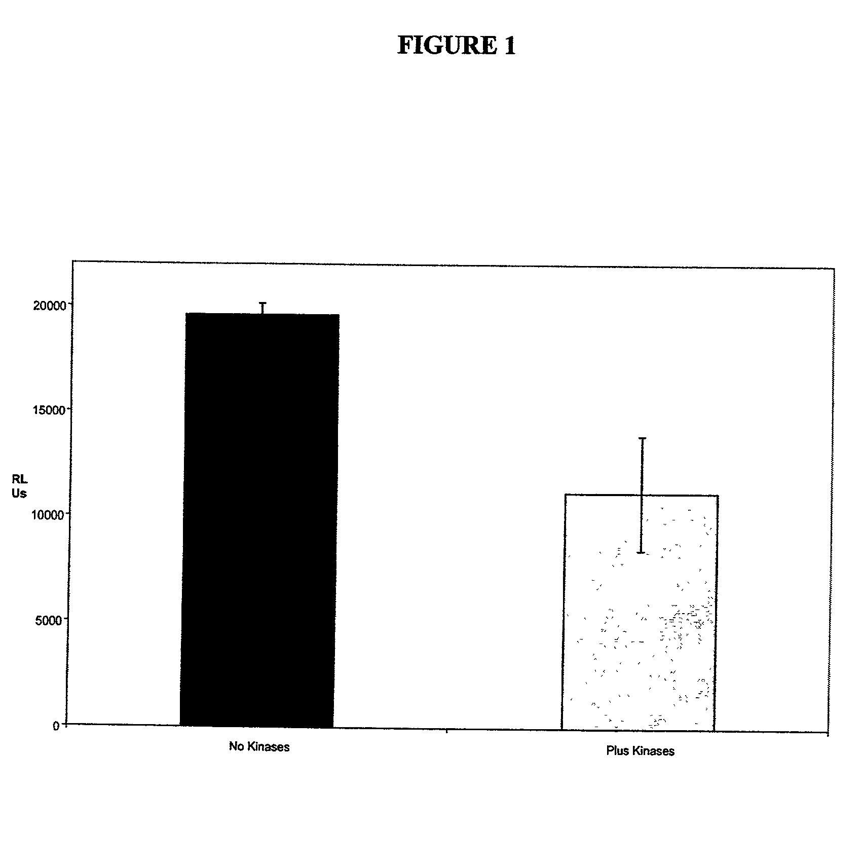
[0100] The first set of experiments was to determine the activity of JNK-1 after the activation of this enzyme by two other kinases, MEKK1 and MKK4. The assay buffer used to activate the enzymes was made up as a 10 times stock (the formulation is shown in appendix 1). The assay was performed as follows, with an initial preparation of a pre-mix for JNK activation.

[0101] The JNK enzyme was used at a stock concentration of 1.234 mg / ml, MEKK1 at 1 mg / ml and MKK4 at 0.28 mg / ml. The activation mixture was added to a polypropylene tube (Sarstedt) with 8.7 .mu.l of JNK, 2.5 .mu.l of MEKK1, 8.4 .mu.l of MKK4, 1 .mu.l of 10 mM ATP (Calbiochem, UK), 1 .mu.l of dithiothreitol (Sigma, UK), 5 .mu.l of 10X assay buffer and 23.4 .mu.l of distilled water. This was incubated overnight at room temperature. To perform the actual assay, the 50 .mu.l of activated concentrate was diluted in 9 mls of JNK assay buffer at the working concentration (i.e. diluted 1:10). Th...

example 2--

Determination of the Activity of MAPK-1 / ERK-1

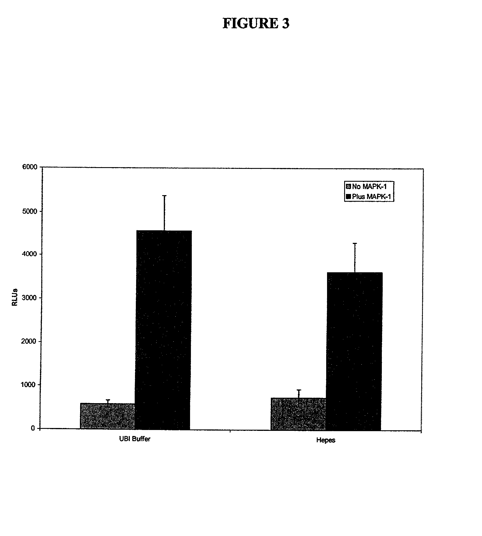
[0104] To confirm that monitoring of kinase activity by the above method of the invention would proceed with other enzymes that cleave the phosphate from ATP, we looked at a number of other kinases which are important in signal transduction and used as targets in drug discovery.

[0105] MAP Kinase-1 / ERK-1 activity was investigated in the presence of myelin basic protein as the phospho-acceptor. This enzyme was supplied in the active form from UBI at a stock concentration of 25 .mu.g in 250 .mu.l. Briefly, 10 .mu.l of assay dilution buffer (UBI, see appendix 1 for formulation) was added to triplicate wells of a white-walled 96 well microtitre plate (Dynex). To this was added 10 .mu.l of MAPK1 and 10 .mu.l of myelin basic protein (UBI, 2 mg / ml), plus 10 .mu.l of ATP cocktail (UBI, for formulation see appendix 1). The plate was sealed and the reaction was allowed to proceed for 10 minutes at 30.degree. C. After this time 110 .mu.l of either as...

example 3--

Determination of the Activity of MAPK-2 / ERK-2

[0107] The above protocol was also used for determining the effect of MAPK-2 / ERK-2 in the presence of the same substrate, myelin basic protein. The MAPK-2 was supplied by UBI at a concentration of 2.5 .mu.g of enzyme in 25 .mu.l of buffer (for formulation see appendix 1), with a specific activity of 662.5 U / mg, where 1U=1 nmole of phosphate incorporated into myelin basic protein. This enzyme was compared with the inactive form, also supplied by UBI at a concentration of 12.5 .mu.g in 50 .mu.l. Both enzymes were diluted in UBI assay dilution buffer to allow for the addition of 25 ng of protein in 10 .mu.l to each well. The myelin basic protein was added as described in the above example, and 200 mM Hepes was used as the buffer added to the wells prior to the addition of the ATP monitoring reagent. The assay was performed in triplicate wells and showed RLUs of 10075.+-.339 for the inactive enzyme, which was very similar to the light output ...


PUM


Abstract

A method for measuring protein kinase activity comprising: (a) providing a first solution comprising ATP and a protein kinase to be tested, and a second solution comprising ATP in the absence of said kinase to be tested; (b) adding a substrate capable of being phosphorylated by the protein kinase to be tested to the first and second solutions of step (a); (c) measuring the concentration of ATP and/or ADP, or the rate of change thereof with respect to time, in each of the reaction mixtures formed in step (b) using a bioluminescence reaction; and (d) using the information about the concentration of ATP and/or ADP to determine the activity of the protein kinase to be tested.

Description

BACKGROUND OF THE INVENTION[0001] 1. Field of the Invention[0002] The present invention relates to methods for detecting protein kinase activity and kits for performing such methods.[0003] 2. Description of Related Art[0004] Protein kinases play crucial roles in the modulation of a wide variety of cellular events. These enzymes act by transferring phosphate residues to certain amino acids in intracellular polypeptides, to bring about the activation of these protein substrates, and set in motion a cascade of activation controlling events including the growth, differentiation and division of cells. Protein kinases have been extensively studied in the field of tumour biology. A lack of controlled activity of kinases in cells is believed to lead to the formation of tumours. The pharmaceutical industry is constantly in search of drugs that target these kinases, to help with the treatment of a wide variety of tumours. There are at least 1200 protein kinases that are involved in the regula...

Claims


Application Information

Patent Timeline
IPC IPC(8): A61P35/00C12Q1/48
CPCG01N2500/04C12Q1/485A61P35/00
Inventor CROUCH, SHARON PATRICIA MARYSLATER, KEVIN JOHN
Owner LONZA GRP
Why Eureka
Try Eureka