Apparatus for performing surgery inside the human retina using fluidic internal limiting membrane (ILM) separation (FILMS)

a technology of internal limiting membrane and apparatus, which is applied in the field of apparatus for performing surgery inside the human retina using fluidic internal limiting membrane (ilm) separation, can solve the problems of loss of fine vision to the level of legal blindness, limiting visual recovery, and legal blindness

Inactive Publication Date: 2003-10-16
MORRIS ROBERT E
View PDF4 Cites 1 Cited by
  • Summary
  • Abstract
  • Description
  • Claims
  • Application Information

AI Technical Summary

Problems solved by technology

This can cause loss of fine vision to the level of legal blindness.
This ILM cellophaning may persist or occur months after seemingly successful removal of EMP, limiting visual recovery.
A macular hole is thought to occur as a result of tangential traction on the retina at the macula, usually leading to legal blindness
This forceps procedure is the most delicate surgical maneuver performed on the human body.
Even minimal ILM surface traction has been increasingly implicated in many forms of maculopathy and often the EMP/ILM layers become, in effect, fused together, not allowing surgical removal of EMP alone.
However, the methods thus far developed for such ILM removal have certain deficiencies.
The method of mechanical pulling tearing away the macular ILM with forceps can cause severe trauma to the macula and the resultant injury can cause ocular damage of equal severity to the problem the surgery is meant to correct.
One problem with the current method of tearing and peeling away the macular ILM is the physical trauma associated with pulling on the ILM until it separates thereby unavoidably stressing the underlying nerve tissue, sometimes causing irreparable nerve damage with worsened vision than may have been present preoperatively.
Accordingly, the surgeon may proceed slowly and carefully but if too slowly the retina may be injured from light toxicity coming from the fiberoptic probe inside the eyeball enabli

Method used


Image

  • Apparatus for performing surgery inside the human retina using fluidic internal limiting membrane (ILM) separation (FILMS)
  • Apparatus for performing surgery inside the human retina using fluidic internal limiting membrane (ILM) separation (FILMS)
  • Apparatus for performing surgery inside the human retina using fluidic internal limiting membrane (ILM) separation (FILMS)

Examples


Embodiment Construction

[0042] It must be noted that as used herein and in the appended claims, the singular forms "a" "and" and "the" include the plural references unless the context clearly dictates otherwise. Thus, for example, reference to "a formulation" includes mixtures of different formulations and reference to "the method of treatment" includes reference to equivalent steps and methods known to those skilled in the art, and so forth.

[0043] Unless defined otherwise, all technical and scientific terms used herein have the same meaning as commonly understood by one of ordinary skill in the art to which this invention belongs. Although any methods and materials similar or equivalent to those described herein can be used in the practice or testing of the invention, the preferred methods and materials are now described. All publications mentioned herein are fully incorporated herein by reference.


PUM


Abstract

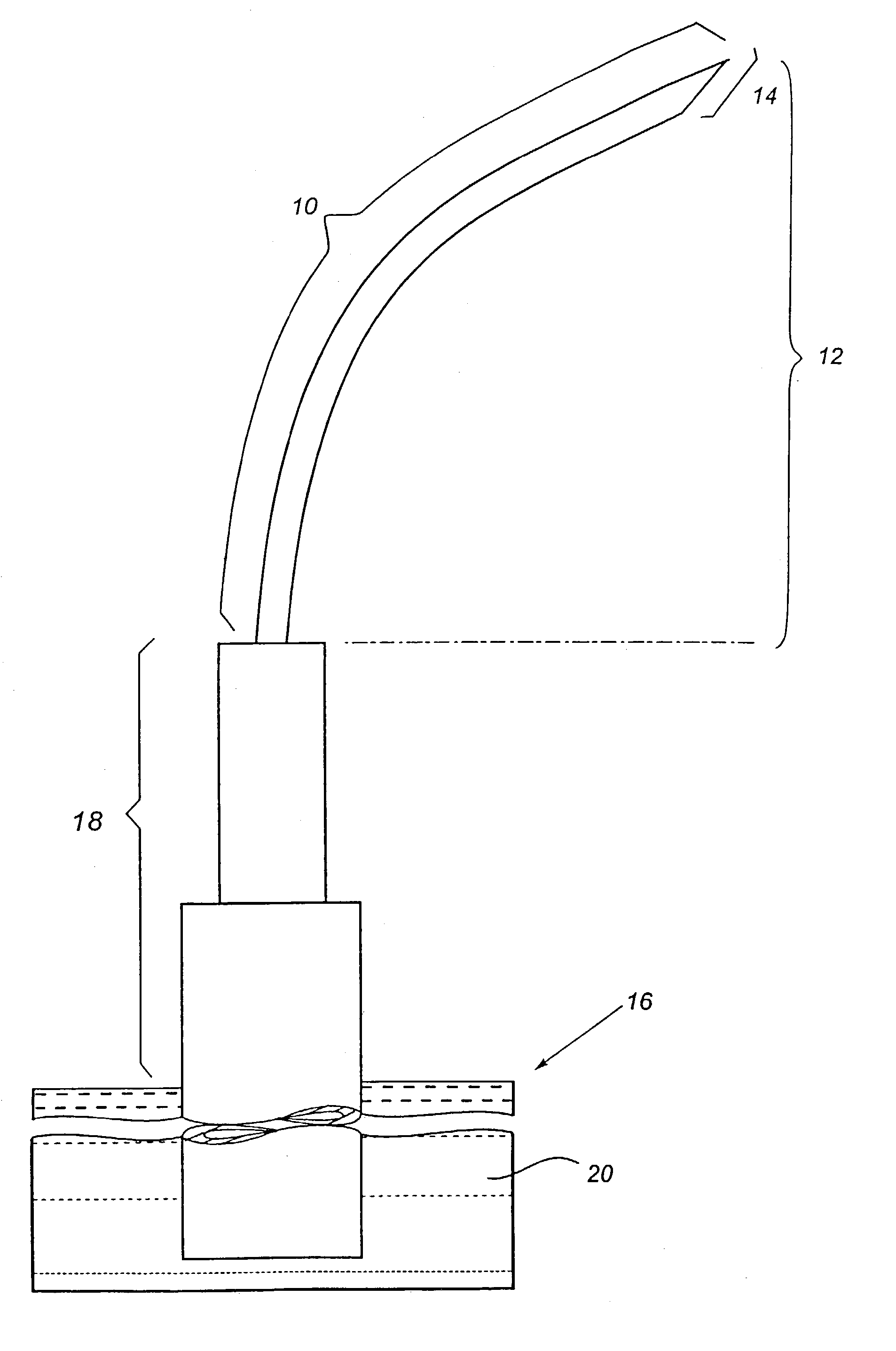
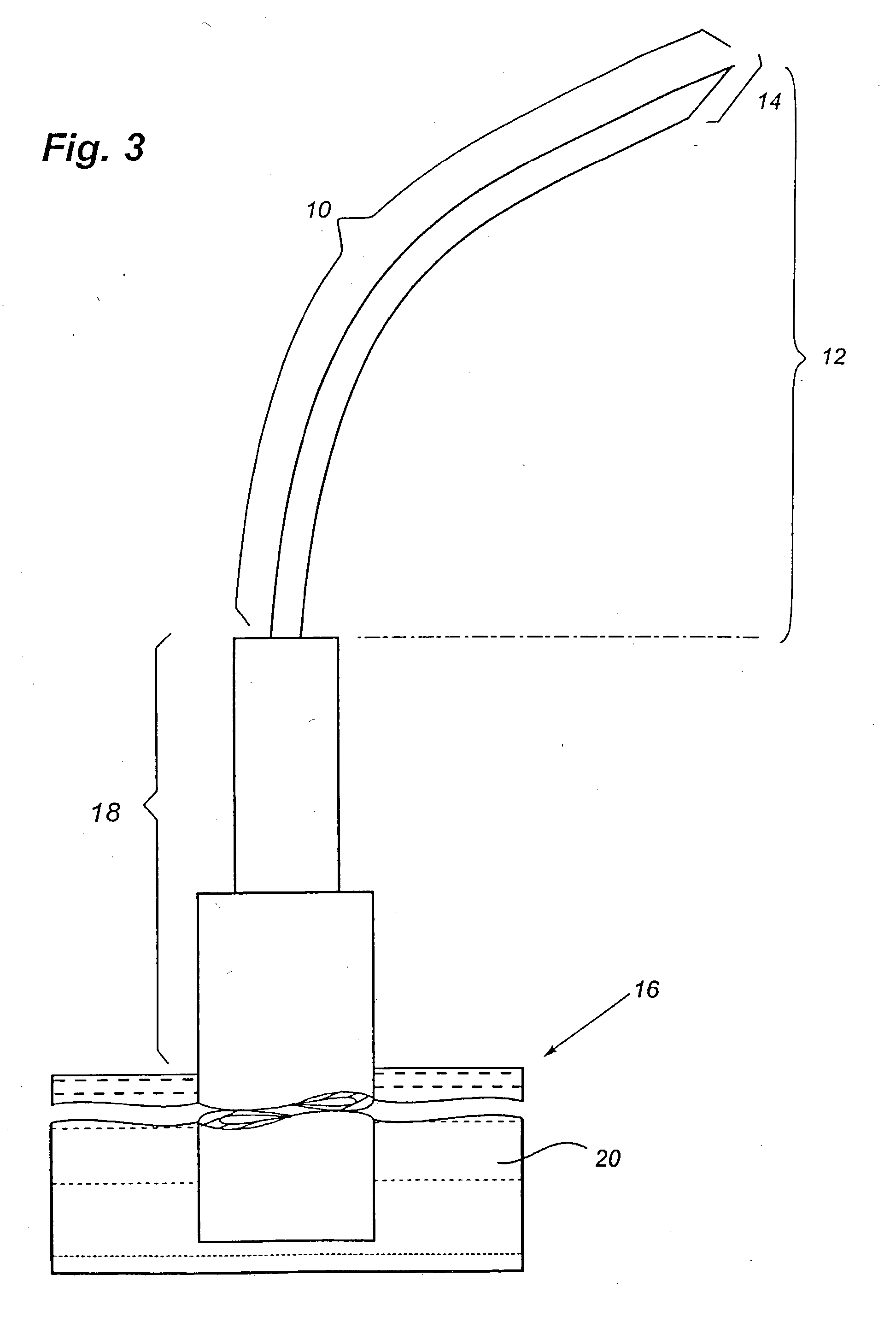
A method and apparatus for performing surgery inside the human retina using fluidic internal limiting membrane separation (FILMS) to remove the internal limiting retinal layer from the neural retinal layer at the macula. The method comprises inserting a hollow microcannula between the retinal internal limiting membrane and the neural retinal layer at or near the macula and injecting a sterile fluid, such as sodium hyaluronate, through said microcannula and thereby raising the macular internal limiting membrane retinal layer away from the neural retina such that it can then be removed by conventional means, while simultaneously smoothing the neural retina by localized pressure tamponad. The apparatus comprises a hollow microcannula having a proximal end a distal end and a distal tip. The distal end is shaped to conform tangentially to the surface of the retina. The distal tip is sharply beveled and adapted to discharge a fluid substance and to easily insert under the internal limiting membrane retinal layer and achieve occlusion of the lumen upon minimal insertion, and is sufficiently microscopic as to not substantially injure the neural retina when introduced under the ILM.

Description

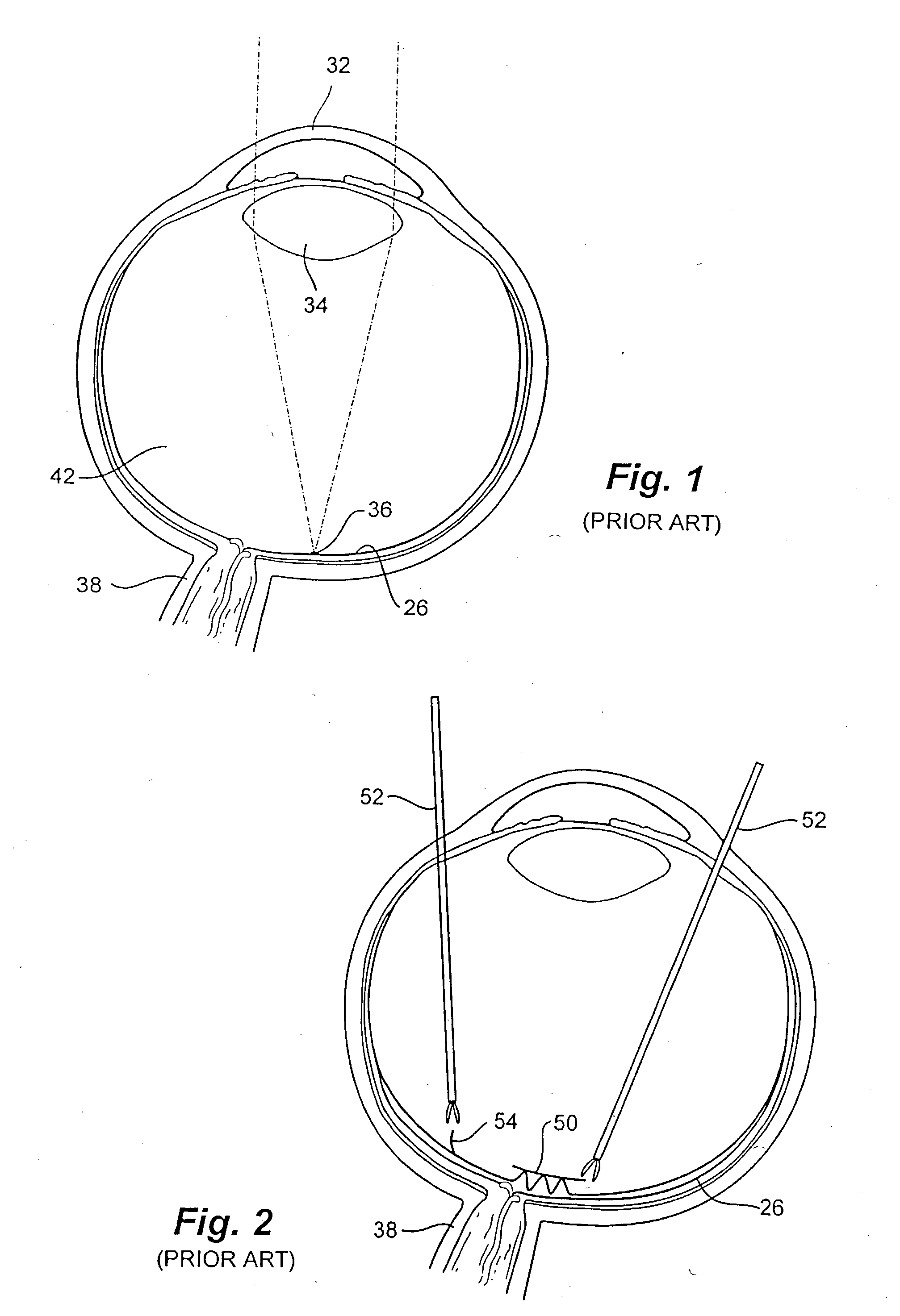
[0001] THIS IS A DIVISON OF U.S. Ser. No. 09 / 111,146 FILED Jul. 6, 1998, THE DISCLOSURE OF WHICH IS HEREIN INCORPORPORATED BY REFERENCE.[0002] 1. Field of the Invention[0003] The present invention is directed generally to medical procedures and, more particularly to a medical procedure for removal of the innermost layer of the human retina (internal limiting membrane) from the underlying neural retina at the center of vision (macula).[0004] 2. BACKGROUND ART[0005] The rays of light entering the eye (FIG. 1) and bearing the pattern of the object being looked upon pass through the cornea 32, the aqueous humor, the pupil, the lens 34, and the vitreous humor, then fall upon the retina 26. The retina is the light sensitive film lining the back two-thirds of the eye. Its appearance is similar to that of wet tissue paper. Its layers consist of the internal limiting membrane (ILM), the neurosensory retina, and the retinal pigment epithelium; the ILM, being innermost, is the retinal border w...

Claims


Application Information

Patent Timeline
IPC IPC(8): A61B17/22A61M1/00A61B17/28A61F9/007A61M25/00
CPCA61M25/00A61M2210/0612A61M2025/0042A61M25/0043A61F9/00736
Inventor MORRIS, ROBERT E.
Owner MORRIS ROBERT E
Why Eureka
Try Eureka