
Human foreskin cells suitable for culturing stem cells

a technology of stem cells and human skin, which is applied in the field of human skin cells suitable for culturing stem cells, can solve the problems of limiting the use of mouse es cells as a model of human development, preventing differentiation of human es cells, and substantially increasing the cost of production from the use of feeder cells

Active Publication Date: 2004-04-08
TECHNION RES & DEV FOUND LTD
View PDF3 Cites 26 Cited by
  • Summary
  • Abstract
  • Description
  • Claims
  • Application Information

AI Technical Summary

Problems solved by technology

Although mouse ES cells facilitate the understanding of developmental processes and genetic diseases, significant differences in primates and mouse development limit the use of mouse ES cells as a model of human development.
However, unlike mouse ES cells, the presence of exogenously added LIF does not prevent differentiation of the human ES cells.
Furthermore, the use of feeder cells substantially increases the cost of production, and makes scale-up of human ES cell culture impractical.
Procedures are not yet developed for completely separating feeder cell components away from embryonic cells prepared in bulk culture.
Thus, the presence of xenogeneic components from the feeder cells complicates their potential use in human therapy.
Although this system uses a better-defined culture conditions, the presence of mouse cells in the culture exposes the human culture to pathogens which restricts their use in cell-based therapy.
However, this culturing method is limited by the high costs of both the matrix and the production of MEF conditioned medium.
In addition, both the matrix and the conditioned medium consist of mouse material, which is basically inconsistent in terms of composition.
However, the major disadvantage of all the abovementioned animal-based xenosupport systems (i.e., MEF with serum and serum replacement, extracellular matrices and conditioned medium) is that they present the risk of animal pathogen cross-transfer to the human ES cells, thus compromising future clinical application.
However, the major disadvantage of using human embryonic fibroblasts or adult fallopian tube epithelial cells as feeder cells is that both of these cell lines have a limited passage ability of only 8-10 times, thereby limiting the ability of a prolonged ES growth period.
While the first are primarily limited by a laborious effort involved in the continuous preparation of fresh feeder layers and the undesired exposure of the human culture to xenogenic components which restrict their future use in therapy, the latter are limited by a shortened culturing period resulting in heterogeneous cultures.
In addition, these human feeder cells are characterized by a relatively long doubling time and a limited culturing period of 7-10 passages which limits their use as a feeder layer.

Method used


Image

  • Human foreskin cells suitable for culturing stem cells
  • Human foreskin cells suitable for culturing stem cells
  • Human foreskin cells suitable for culturing stem cells

Examples


example 1

[0122] Foreskin--Derived Cell Lines Suitable for Growing ES Cells

[0123] Materials and Experimental Methods

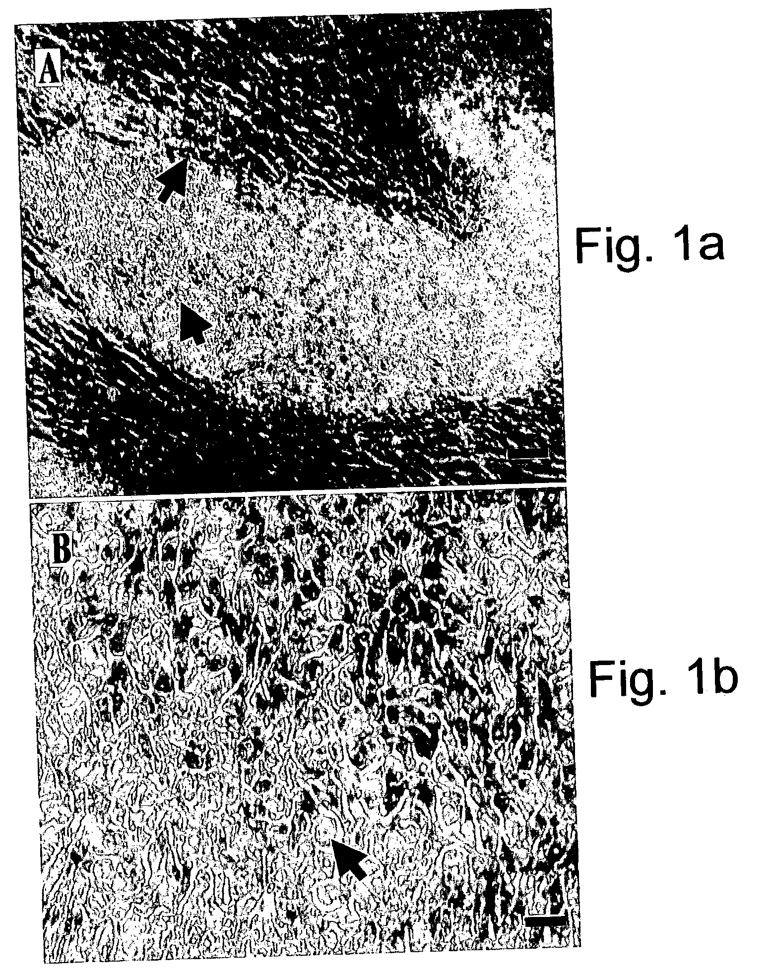
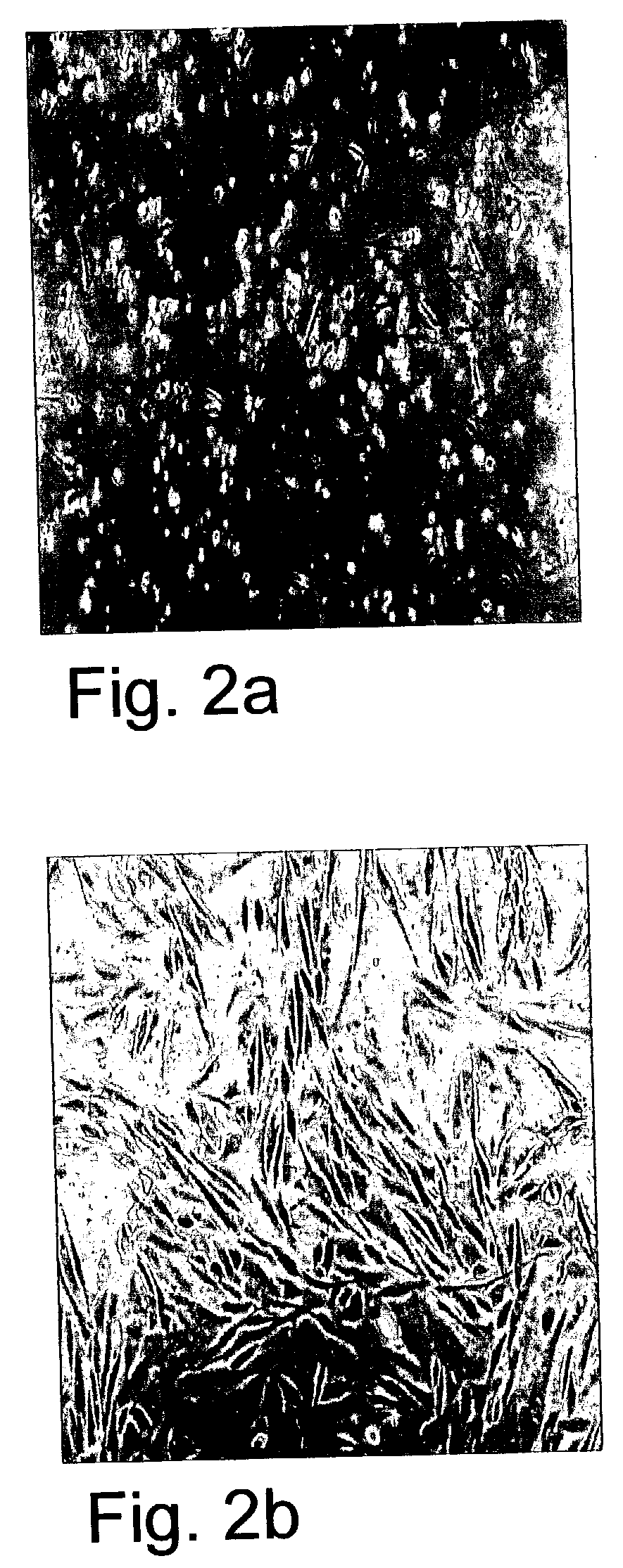
[0124] Establishment of foreskin feeder cell lines--Foreskin tissue was obtained by parental informed consent from 8 to 14 day old normal males. Tissue was washed in sterile PBS (Invitrogen, Grand island, N.Y., USA), minced via scissors and dissociated to single cells by incubation with Trypsin-EDTA (0.5% Trypsin, 5.3 mM EDTA, Invitrogen, Grand Island, N.Y., USA) for 20-40 min. Resulting single cell suspensions were grown in a culture medium including 80% Dulbecco's modified Eagle's medium (DMEM) containing high glucose concentration and no pyruvate (Invitrogen, Grand island, N.Y., USA), 2 mM L-glutamine, 0.1 mM .beta.-mercaptoethanol, and 1% non-essential amino acid stock (all purchased from Invitrogen, Grand island, N.Y., USA products). Foreskin medium was supplemented with serum or serum replacement as follows: Growth medium for foreskin cell lines F3, F7 and F8 was supplemen...

example 2

[0134] Foreskin-Derived Feeder Cell Lines Support Growth of Phenotypically Consistent ES Cells

[0135] Materials and Experimental Methods

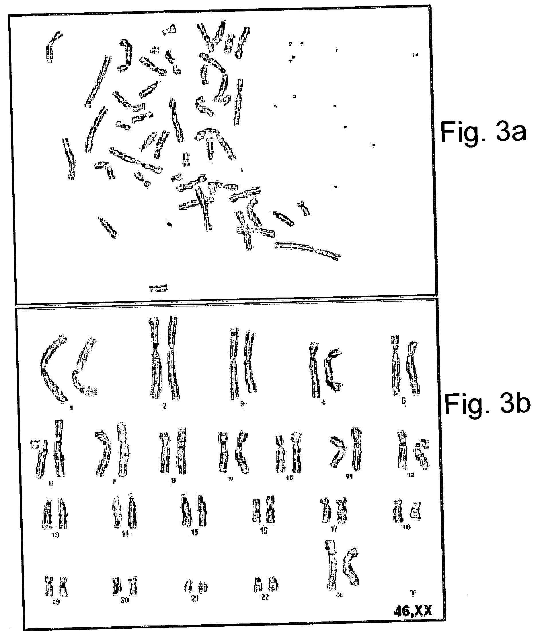
[0136] Karyotype analysis--ES cells metaphases were blocked using colcemid (KaryoMax colcemid solution, Invitrogen, Grand island, N.Y., USA) and nuclear membranes were lysed in an hypotonic solution according to standard protocols (International System for Human Cytogenetic Nomenclature, ISCN). G-banding of chromosomes was performed according to manufacturer's instructions (Giemsa, Merck). Karyotypes of at least 50 cells per sample were analyzed and reported according to the ISCN.

[0137] Immunohistochemistry--Cells were fixed for 20 min in 4% paraformaldehyde, blocked for 15 min in 2% normal goat serum in PBS (Biological Industries, Beth Haemek, Israel) and incubated for overnight at 4.degree. C. with 1:50 dilutions of SSEA1, SSEA3, SSEA4, TRA-60, TRA-81 mouse anti-human antibodies, provided by Prof. P Andrews the University of Sheffield, England. Cel...

example 3

[0145] Foreskin-Derived Feeder Cells Support Growth of Functional ES Cells

[0146] Materials and Experimental Methods:

[0147] Formation of embryoid bodies (EBs) from human ES cells--Human ES cells grown on foreskin derived feeder layers were removed from the 6-well plate (60 cm.sup.2) co-culture by Type IV Collagenase (1 mg / ml) and were further dissociated into small clamps using 1000 .mu.l Gilson pipette tips. Thereafter, dissociated cells were cultured in 58 mm Petri dishes (Greiner, Germany) in a medium consisting of 80% Ko-DMEM, supplemented with 20% fetal bovine serum defined (FBSd, HyClone, Utah, USA), 1 mM L-glutamine, 0.1 mM .beta.-mercaptoethanol, and 1% non-essential amino acid stock. Unless otherwise noted all were purchased from Gibco Invitrogen corporation, USA. Formation of EBs was examined following one month in suspension.

[0148] Reverse transcriptase (RT) coupled PCR--Total RNA was isolated from either undifferentiated human ES cells or from one-month-old EBs using Tri-...


PUM


Abstract

A cell culture comprising human foreskin cells, the human foreskin cells being capable of maintaining stem cells in an undifferentiated state when co-cultured therewith.

Description

[0001] This application claims the benefit of priority from U.S. Provisional Patent Application No. 60 / 416,242, filed Oct. 7, 2002.FIELD AND BACKGROUND OF THE INVENTION[0002] The present invention relates to human foreskin cells, which are capable of maintaining stem cells in an undifferentiated state in culture.[0003] Embryonic stem (ES) cells are derived from the inner cell mass (ICM) of the mammalian blastocyst (Evans & Kaufman 1981; Martin 1981). These cells are pluripotent thus capable of developing into any organ or tissue type and even a complete embryo. When cultured in suspension ES cells aggregate and differentiate into simple embryoid bodies (EBs), however once cultured under suitable conditions (as further described hereinbelow), true ES cells are capable of indefinite proliferation in vitro in an undifferentiated state; maintaining a normal karyotype through prolonged culture; and maintaining the potential to differentiate to derivatives of all three embryonic germ laye...

Claims


Application Information

Patent Timeline
IPC IPC(8): C12N5/0735C12N5/074
CPCC12N5/0606C12N5/0611C12N2506/02C12N2502/13C12N2533/90C12N5/0043C12N5/0056C12N2533/50C12N2500/00C12N5/0031C12N5/0603C12N5/0665C12N5/0695C12N5/0696C12N2500/99C12N2502/1323
Inventor AMIT, MICHALITSKOVITZ-ELDOR, JOSEPH
Owner TECHNION RES & DEV FOUND LTD
Learn More