Rapid immunoassay of anthrax protective antigen in vaccine cultures and bodily fluids by fluorescence polarization

Inactive Publication Date: 2004-11-25
THE UNITED STATES OF AMERICA AS REPRESENTED BY THE SECRETARY OF THE NAVY
2 Cites 12 Cited by

AI-Extracted Technical Summary

Problems solved by technology

Saliva presents several challenges for diagnostics: limited reference values have been published and the standardization of sample collection has fallen behind serum (13).
However, despite the clear advantages of FP in diagnostics, only a limited number of non-commercial laboratories have adopted FP for detection of antibody in serum samples (33-35).
ELISA methods, however, are inadequate due to either low sensitivity or erroneous results caused by interference effects by media constituents.
SDS-PAGE methods are inadequate because they do not give real time information such that a cultu...

Method used

[0041] Quantitative, real-time monitoring of growing bacterial cultures is necessary to ensure maximum production of important bacterial proteins in vaccine production. Bacillus anthracis is of ...

Abstract

The inventive subject matter relates to a competitive method for estimating the concentration in a sample of a Bacillus anthracis protein or antibody thereof selected from the group consisting of protective antigen (PA), lethal factor (LF) and edema factor (EF). The method may employ the use of Fluorescence Polarization, FLT or FRET. The competitive methods are capable of detecting a target protein within 5 minutes within the range of 0.1 to 10.0 nM. The methods provide for the rapid detection and quantitation of bacteria, bacterial antigen or antibody in culture media or broth of growing cultures of bacteria, including B. anthracis by fluorescent methods.

Application Domain

Microbiological testing/measurementPeptide preparation methods +3

Technology Topic

Quantitative cultureEdema +18

Image

  • Rapid immunoassay of anthrax protective antigen in vaccine cultures and bodily fluids by fluorescence polarization

Examples

  • Experimental program(1)

Example

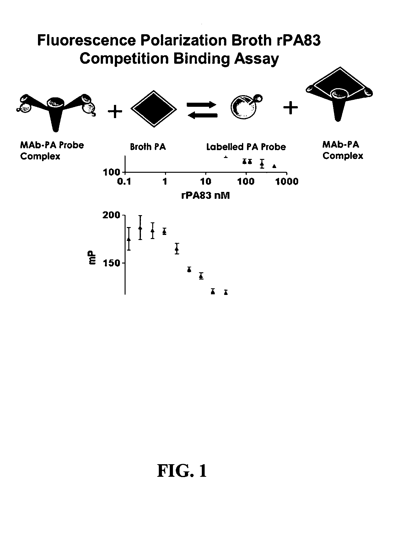
Competitive FP Assay for Measuring Broth Protective Antigen, Lethal Factor and Endema Factor of Bacillus anthracis
[0041] Quantitative, real-time monitoring of growing bacterial cultures is necessary to ensure maximum production of important bacterial proteins in vaccine production. Bacillus anthracis is of particular importance due to its potential public health and military focus as a potential bioterrorism weapon. Monitoring of growing cultures of B. anthracis is conducted by the following steps:
[0042] a) Add 10 .mu.l of labeled competitor peptide (e.g. FITC labeled PA, lethal factor or edema factor) in 1 ml buffer;
[0043] b) Read blank;
[0044] c) Add 20 .mu.l of monoclonal antibody to target protein in said 1 ml buffer;
[0045] d) Incubate for 2 minutes;
[0046] e) Read the FP of the tracer;
[0047] f) Add 10 .mu.l of culture broth to said 1 ml buffer in step d);
[0048] g) Read decrease in mP. The decrease in mP is proportional to the PA concentration.
[0049] Culture broths of B. anthracis contain protective antigen concentrations up to 5 ug/ml. Therefore the assayable range of protective antigen required for FP assay is similar to that for ELISA assay. Using standard sandwich ELISA techniques, recombinant PA reports approximately 0.01 OD per 1 ng PA at 405 nm using horse radish peroxidase substrate conjugated antibodies in a range of 1.5-200 ng protective antigen. An example standard ELISA was used to estimate protective antigen in serial dilutions of buffer from 25 nM (2 ug/ml) down to 0.2 nM (16 ng/ml).
[0050] Similar results are expected in competitive fluorescence polarization as immunoassay techniques (see FIG. 1). Combining an fluorescent dye-labeled protective antigen probe at about 1 nM (labeled PA probe) and a monoclonal (MAb) or polyclonal (PAb) antibody at an adequate dilution (usually between 1:25-1:250K) would report numbers around 200 mP (left side of equation and left side of graph as shown in FIG. 1). Culture broth would be added as unlabeled competitor (broth PA) in a range of 0.01-0.1 ml depending on how long the culture had grown causing dissociation of the labeled probe from the antibody. Dissociation of the labeled probe from the antibody in the presence of increasing amounts of broth PA results in increasing amounts of free labeled probe and decreasing mP readings that are proportional to the concentration of protective antigen in the culture broth. (Right side of the equation and right side of the graph). At full growth, the assay mixture would require 0.01 ml broth (5 ug/ml protective antigen, about 6 nM) while partial growth would require proportionately more broth up to 0.1 ml to estimate a similar concentration. Other optional reagents may be used to suppress intrinsic culture broth fluorescence. cl EXAMPLE 2
Determining the Concentration of Recombinant or Native Protective Antigen (PA) of Bacillus anthracis Antigen in Fermentation Broth
[0051] Overlapping 15 and 25 mer peptide sequences were synthesized from the amino acid region 1-764 of full-length PA using Fmoc (9-fluorenylmethoxycarbonyl) chemistry including a final step to attach an N-terminal 6-carboxyfluorescein (FAM). Enzymatic digests of fluorescein-6-isothiocyanate (FITC isomer II) labeled full length PA were made with trypsin, pepsin and chymotrypsin. These peptides were screened for specific antibody binding by immune sera. Using Western Blot, ELISA and dot-blot analysis, a 10-mer portion of domain 4 of the COOH region of PA was suitable as an FP probe, a 5-6 kDa portion of domain 3 and domain 4 was suitable as an FP probe, a 10 kDa portion of domain 3 and 4 was suitable as a FP probe, a 5-6 kDa portion of domain 1 and 2 was suitable as a FP probe, and a 10 kDa portion of domain 1 and 2 was suitable as a FP probe
[0052] Fermentation broth cultures of Bacillus anthracis (vaccine strain V770-NP1-R) containing PA were produced. Culture media is then mixed with antibody and competitive probe. Incubation times was less than 30 seconds for cultures nearing the stationary phase of growth and for less than 4 minutes for newly seeded cultures (and therefore low concentration of PA). At 1 nM probe concentration, the blank subtracted fluorescence polarization reading is determined to be quantifiable if MP decreased by at least 10 mP.
References
[0053] 1. Anonymous: Biography on Saliva; Library of Congress Bibliography Section Science & Technology Division; Office of Naval Research Report ACR-48, Washington, D.C., 1960. Library of Congress.
[0054] 2. Kraus F, Konno J: The salivary secretion of antibody, Alabama J Med Sci 1965; 2:15-22.
[0055] 3. Hirschman J D, Kresge J A: Synthesis of a symposium: innovative non- or minimally-invasive technologies for monitoring health and nutritional status in mothers and young children, J Nutr 2001; 131:1643S-5S.
[0056] 4. Hofman L F: Human saliva as a diagnostic specimen, J Nutr 2001; 131:1621S-5S.
[0057] 5. Tenovuo J: Human Saliva: Clinical Chemistry and Microbiology, 256. Vol. 1, Boca Raton, Fla., CRC Press, 1989.
[0058] 6. Proceedings: Saliva in health and disease. Proceedings of the 16th International Conference on Oral Biology. Apr. 9-12, 2000. Chantilly, Va., USA. Proceedings, Adv Dent Res 2000; 14:5-102.
[0059] 7. Kaufman E, Lamster I B: Analysis of saliva for periodontal diagnosis--a review, J Clin Periodontol 2000; 27:453-65.
[0060] 8. Streckfus C F, Bigler L R: Saliva as a diagnostic fluid, Oral Dis 2002; 8:69-76.
[0061] 9. Rossomando E F, Kousvelari E, Janicki B W, Tabak L A: Improvement of oral health in the postgenomic era: opportunities for government/industry partnerships, Compend Contin Educ Dent 2001; 22:570-2, 574.
[0062] 10. Tabak L A: A revolution in biomedical assessment: the development of salivary diagnostics, J Dent Educ 2001; 65:1335-9.
[0063] 11. Cordeiro M L, Turpin C S, McAdams S A: A comparative study of saliva and OraSure oral fluid, Ann N Y Acad Sci 1993; 694:330-1.
[0064] 12. Rehak N N, Cecco S A, Csako G: Biochemical composition and electrolyte balance of "unstimulated" whole human saliva, Clin Chem Lab Med 2000; 38:335-43.
[0065] 13. Soderling E: Practical Aspects of Salivary Analysis, Human Saliva: Clinical Chemistry and Microbiology, 1-24. Vol. 1, Boca Raton, CRC Press, 1989.
[0066] 14. Seymour E: Salivary sampling device and sample adequacy system: Pat. No. 5,393,496. Patent and Trademark Office, United States, Salivary Diagnostic Systems, Vancouver, Wash., 1995.
[0067] 15. Goldstein A S, Gadojdea S, Zogg D F: Oral collection device and method for immunoassay: Pat. No. 5,335,673. Patent and Trademark Office, United States, Epitope, Inc., Beaverton, Oreg., 1994.
[0068] 16. McKie A, Vyse A, Maple C: Novel methods for the detection of microbial antibodies in oral fluid, Lancet Infect Dis 2002; 2:18-24.
[0069] 17. Marcus E R, Jooste C P, Driver H S, Hattingh J: The quantification of individual proteins in crevicular gingival fluid, J Periodontal Res 1985; 20:444-9.
[0070] 18. Burke J C, Evans C A, Crosby T R, Mednieks M I: Expression of secretory proteins in oral fluid after orthodontic tooth movement, Am J Orthod Dentofacial Orthop 2002; 121:310-5.
[0071] 19. Slots Jr, Taubman M A: Contemporary oral microbiology and immunology, 649, St. Louis, Mosby-Year Book, 1991.
[0072] 20. Thieme T R, Goldstein A S, Piacentini S C, Klimkow N M: Oral Collection Device and Kit: Pat. No. 5,830,410. Patent and Trademark Office, United States, Epitope, Inc., Beaverton, Oreg., 1998.
[0073] 21. Cimasoni G: The crevicular fluid, Monographs in oral science; Vol. 3, 122, Basel, New York,, S. Karger, 1974.
[0074] 22. Lehner T: The Borderland between caries and periodontal disease : Proceedings of a conference sponsored by the Royal Society of Medicine, 28 Feb. 1977, London, New York, 1977. Academic Press, Grune & Stratton.
[0075] 23. Steenhuysen J: Orasure, Abbott agree to co-distribute HIV test. Reuters, Chicago, 2002.
[0076] 24. Roitt I M, Lehner T: Immunology of Oral Diseases, pp. 464, Oxford, Blackwell Scientific Publications, 1981.
[0077] 25. Gallo D, George J R, Fitchen J H, Goldstein A S, Hindahl M S: Evaluation of a system using oral mucosal transudate for HIV-1 antibody screening and confirmatory testing. OraSure HIV Clinical Trials Group, JAMA 1997; 277:254-8.
[0078] 26. Thieme T, Piacentini S, Davidson S, Steingart K: Determination of measles, mumps, and rubella immunization status using oral fluid samples, JAMA 1994; 272:219-21.
[0079] 27. Bull A R, Kimmance K J, Parry J V, Perry K R: Investigation of an outbreak of hepatitis A simplified by salivary antibody testing, Epidemiol Infect 1989; 103:371-6.
[0080] 28. Parry J V, Perry K R, Panday S, Mortimer P P: Diagnosis of hepatitis A and B by testing saliva, Journal Med Virol Aug 1989; 28:255-60.
[0081] 29. Sherman K E, Creager R L, O'Brien J, Sargent S, Piacentini S, Thieme T: The use of oral fluid for hepatitis C antibody screening, Am J Gastroenterol 1994; 89:2025-7.
[0082] 30. Patel P, Mendall M, Khulusi S, et al.: Salivary antibodies to Helicobacter pylori: screening dyspeptic patients before endoscopy, Lancet 1994; 344:511-4.
[0083] 31. Parry J V, Perry K R, Mortimer P P: Sensitive assays for viral antibodies in saliva: an alternative to tests on serum, Lancet 1987; 2:72-5.
[0084] 32. Barros M, Duarte Neto A N, Pereira V R, et al.: Evaluation of oral mucosal transudate for immunodiagnosis of Chagas' disease, Rev Inst Med Trop Sao Paulo 1999; 41:265-6.
[0085] 33. Tencza, S., Islam, K., Kalia, V., Nasir, M., Jolley, M., Montelaro, R. 2000. Development of a fluorescence polarization-based diagnostic assay for equine infectious anemia virus. J. Clin Microbiol. 38: 1854-9.
[0086] 34. Lin, M., Sugden, E., Jolley, M., Stilwell, K. 1996. Modification of the Mycobacterium bovis extracellular protein MPB70 with fluorescein by fluorescence polarization. Clin Diag. Lab Immunol. 3: 438-43.
[0087] 35. Nielsen K, Gall D, Jolley M, et al.. 1996. A homogeneous fluorescence polarization assay for detection of antibody to Brucella abortus, J Inmunolog Methods 195:161-8.
[0088] 36. Adams, C, Boge, A. Gaudet L. 2003. HTS Platform for Determining Enzyme Activity Assay Tutorial. Genetic Engineering News 23(20) 38-39.
[0089] 37. Gallo, D., George, J. R., Fitchen, J. H., Goldstein, A. S., Hindahl, M. S., 1997. Evaluation of a system using oral mucosal transudate for HIV-1 antibody screening and confirmatory testing. OraSure HIV Clinical Trials Group, JAMA 277: 254-8.
[0090] 38. Thieme, T., Piacentini, S., Davidson, S., Steingart, K. 1994. Determination of measles, mumps and rubella immunization status using oral fluid samples. JAMA 272: 219-21.
[0091] 39. Bull, A. R., Kimmance, K. J., Parry, J. V., Perry, K. R. 1989. Investigation of an outbreak of hepatitis A simplified by salivary antibody testing. Epidemiol. Infect. 103: 371-6.
[0092] 40. Parry, J. V., Perry, K. R., Panday, S., Mortimer, P. P. 1989. Diagnosis of hepatitis A and B by testing saliva. Journal Med. Virol. 28 (Aug): 255-60.
[0093] 41. Sherman, K. E., Creager, R. L., O'Brien, J., Sargent, S., Piacentini, S., Thierme, T. 1994. The use of oral fluid for hepatitis C antibody screening. Am J. Gastrogenterol. 89: 2025-7.
[0094] 42. Patel, P., Mendall, M., Khulusi, S., Molineaux, N., Levy, J., Maxwell, J. D., Northfield, T. S. 1994. Salivary antibodies to Helicobacter pylori: screening dyspeptic patients before endoscopy. Lancet 344: 511-514.
[0095] 43. Parry, J. V., Perry, K. R., Mortimer, P. P. 1987. Sensitive assays for viral antibodies in saliva: an alternative t tests on serum. Lancet 2: 72-5.
[0096] 44. Rossomando, E. F., Kousvelari, E., Janicki, B. W., Tabak, L. A. 2001. Improvement of oral health in th postgenomic era: opportunities for government/industry partnerships, Compend. Contin. Educ. Dent. 22: 570-4.
[0097] 45. Tabak, L. A. 2001. A revolution in biomedical assessment: the development of salivary diagnostics. J. Dent Educ. 65: 1335-9.
[0098] 46. Gunderson, E. K., Garland, C., Hourani, L. L. 1995. Infectious disease rates in the U.S. Navy, 1980 to 1995. Mil. Med. 166: 544-9.
[0099] 47. Behr, M. A. 2004. Tuberculosis due to multiple strains: a concern for the patient? A concern for tuberculosis control? Am. J. Respir. Crit. Care. Med. 169 (5): 554-5.
[0100] 48. Sellman, B., Mourez, M., Collier, R. 2001. Dominant-negative mutants of a toxin subunit: An approach to therapy of anthrax. Science 292: 695-7.
[0101] 49. Petosa, D., Collier, R., Klimpel, K., Leppla, S., Liddington, R. 1997. Crystal structure of the anthrax toxin protective antigen. Nature 385: 833-8.
[0102] 50. Little, S. F., Novak, J., Lowe, J., Singh, Y., Klimpel, K. R., Lingerding, B. C., and Friedlander, A. M. 1996. Characterization of lethal factor binding and cell receptor binding domains of protective antigen of Bacillus anthracis protective antigen. Microbiology 142: 707-15.
[0103] 51. Brossier, F., Sirard, J-C., Guidi-Rontani, C., Duflot, E., Mock., M. 1999. Functional analysis of the carboxy-terminal domain of Bacillus anthracis protective antigen. Infect. Immun. 67: 964-7.
[0104] 52. Brossier, F., Weber-Levy, M., Mock, M., Sirard, J. 2000. Role of toxin functional domains in anthrax pathogenesis. Infect. Immun. 68: 1781-6.
[0105] 53. Mogridge, J., Moourez, M., Collier, R. 2001. Involvement of domain 3 in oligomerization by the protective antigen moiety of anthrax toxin. J. Bacteriol. 183: 2111-6.
[0106] 54. Brachman, P., Gold, H., Plotkin, S., Fekety, F., Werrin, M., Ingraham, N. 1962. Field evaluation of a human anthrax vaccine. Am. J. Public Health 52: 632-45.
[0107] 55. Fellows, P., Linscott, M., Ivins, B., Pitt, M. L. M., Rossi, C. A., Gibbs, P. H., and Friedlander, A. M. 2001. Efficacy of a human anthrax vaccine in guinea pigs, rabbits, and rhesus macaques against challenge by Bacillus anthracis isolates of diverse geographical origin. Vaccine 19: 3241-7.
[0108] 56. Turnbull, P., Broster, M., Carman, J., Manchee, R., Melling, J. 1986. Development of antibodies to protective antigen and lethal factor components of anthrax toxin in humans and guinea pigs and their relevance to protective immunity. Infect Immun. 52: 356-63.
[0109] 57. Kowski T, Wu J: Fluorescence polarization is a useful technology for reagent reduction in assay miniaturization, Comb Chem High Throughput Screen 2000; 3:437-44.
[0110] 58. ONR: Future Naval Capabilities: Warfighter Protection, Washington, D.C., Department of the Navy: Science and Technology Lead, 1992.
[0111] 59. Terpetschnig E, Szmacinski H, Lakowicz J: Fluorescence polarization immunoassay of a high-molecular-weight antigen based on a long-lifetime Ru-ligand complex, Anal Biochem 1995; 227:140-7.
[0112] 60. Tencza S, Islam K, Kalia V, Nasir M, Jolley M, Montelaro R: Development of a fluorescence polarization-based diagnostic assay for equine infectious anemia virus, J Clin Microbiol 2000; 38:1854-9.
[0113] 61. Schade S, Jolley M: Fluorescence polarization assays of enzymes and substrates thereof: Pat. No. 5,804,395. Patent and Trademark Office, United States, Secretary of the Navy, 1998.
[0114] 62. Nasir M, Jolley M: Fluorescence polarization: An analytical tool for immunoassay and drug discovery, Comb Chem High Throughput Screen 1999; 2:177-90.
[0115] 63. Nasir M, Jolley M: Fluorescence polarization: Stretching it to its limits. A special edition on fluorescence polarization., Comb Chem High Throughput Screen 2002; in press.
[0116] 64. Jolley M: Fluorescence polarization immunoassay for the determination of therapeutic drug levels in human plasma, J Anal Toxicology 1981; 5:236-40.
[0117] 65. Lin M, Sugden E, Jolley M, Stilwell K: Modification of the Mycobacterium bovis extracellular protein MPB70 with fluorescein by fluorescence polarization, Clin Diag Lab Immunol 1996; 3:438-43.
[0118] 66. Nielsen K, Gall D, Jolley M, et al.: A homogeneous fluorescence polarization assay for detection of antibody to Brucella abortus, J Immunolog Methods 1996; 195:161-8.
[0119] 67. Cui, H. H., Valdez, J. G., Steinkamp, J. A., Crissman, H. A. 2003. Fluorescence lifetime-based discrimination and quantification of cellular DNA and RNA with phase-sensitive flow cytometry. Cytometry 52A(1): 46-55.

PUM

PropertyMeasurementUnit
Fraction0.96 ~ 0.99fraction
Time15.0s
Time30.0s

Description & Claims & Application Information

We can also present the details of the Description, Claims and Application information to help users get a comprehensive understanding of the technical details of the patent, such as background art, summary of invention, brief description of drawings, description of embodiments, and other original content. On the other hand, users can also determine the specific scope of protection of the technology through the list of claims; as well as understand the changes in the life cycle of the technology with the presentation of the patent timeline. Login to view more.
Why Eureka
Try Eureka