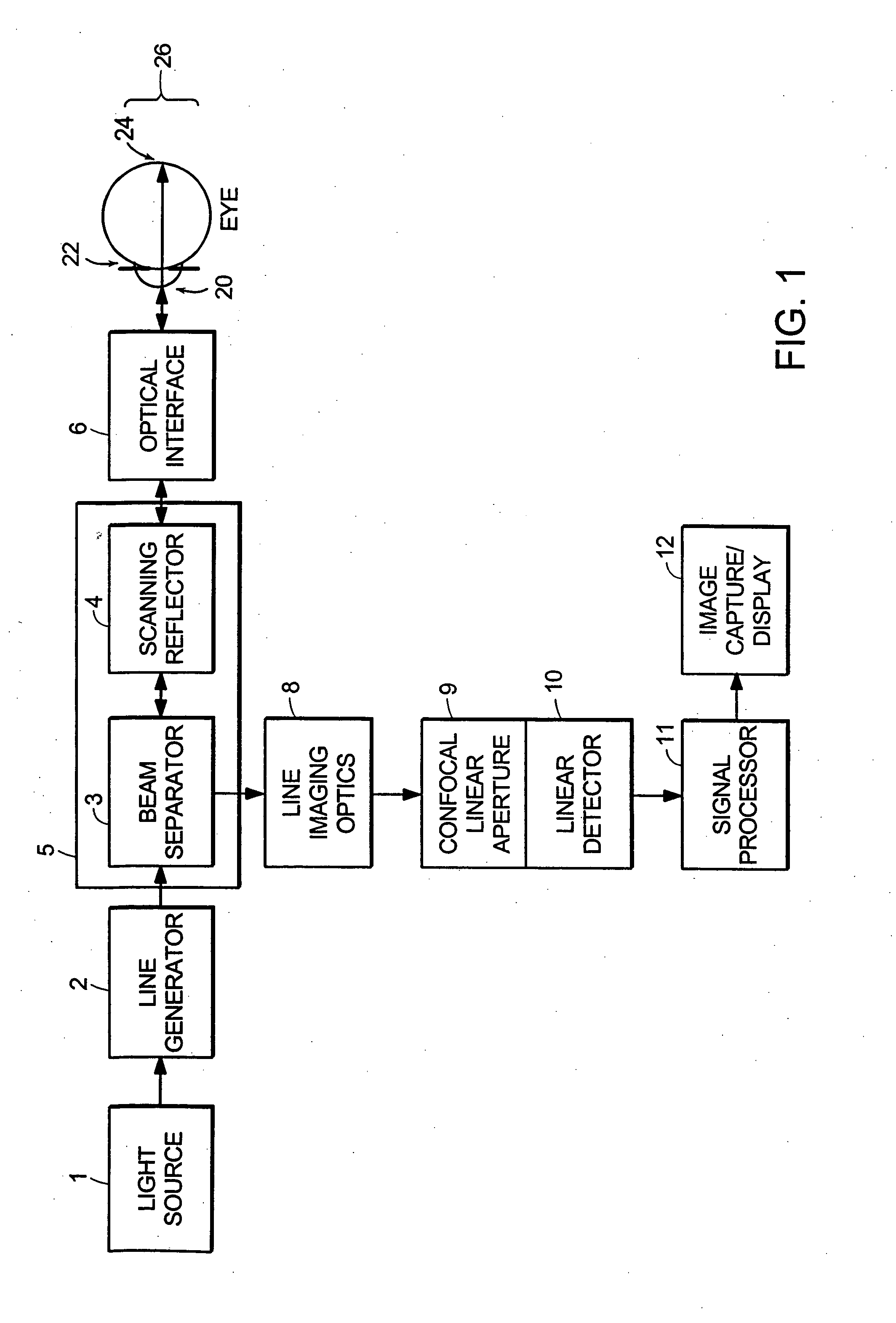
Line-scan laser ophthalmoscope

a laser ophthalmoscope and line-scan technology, applied in the field of eye examination systems and methods, can solve problems such as patient discomfort, and achieve the effects of less expensive, compact design, and significant confocal advantage in image clarity and contras

Inactive Publication Date: 2005-01-20
PHYSICAL SCI
View PDF2 Cites 48 Cited by
  • Summary
  • Abstract
  • Description
  • Claims
  • Application Information

AI Technical Summary

Benefits of technology

The line-scanning laser ophthalmoscope (LSLO) of the invention has a significant confocal advantage in image clarity and contrast, and depth of penetration at the ocular fundus compared with conventional digital fundus photography. The LSLO has features not currently available in commercial SLOs, and is less expensive. The hand-held digital LSLO has proven that high quality, non-mydriatic (e.g., undilated pupil), line-confocal retinal images and stereo pairs can be obtained with a simple, compact design with fewer moving parts and components than current SLO systems. In one embodiment, the 
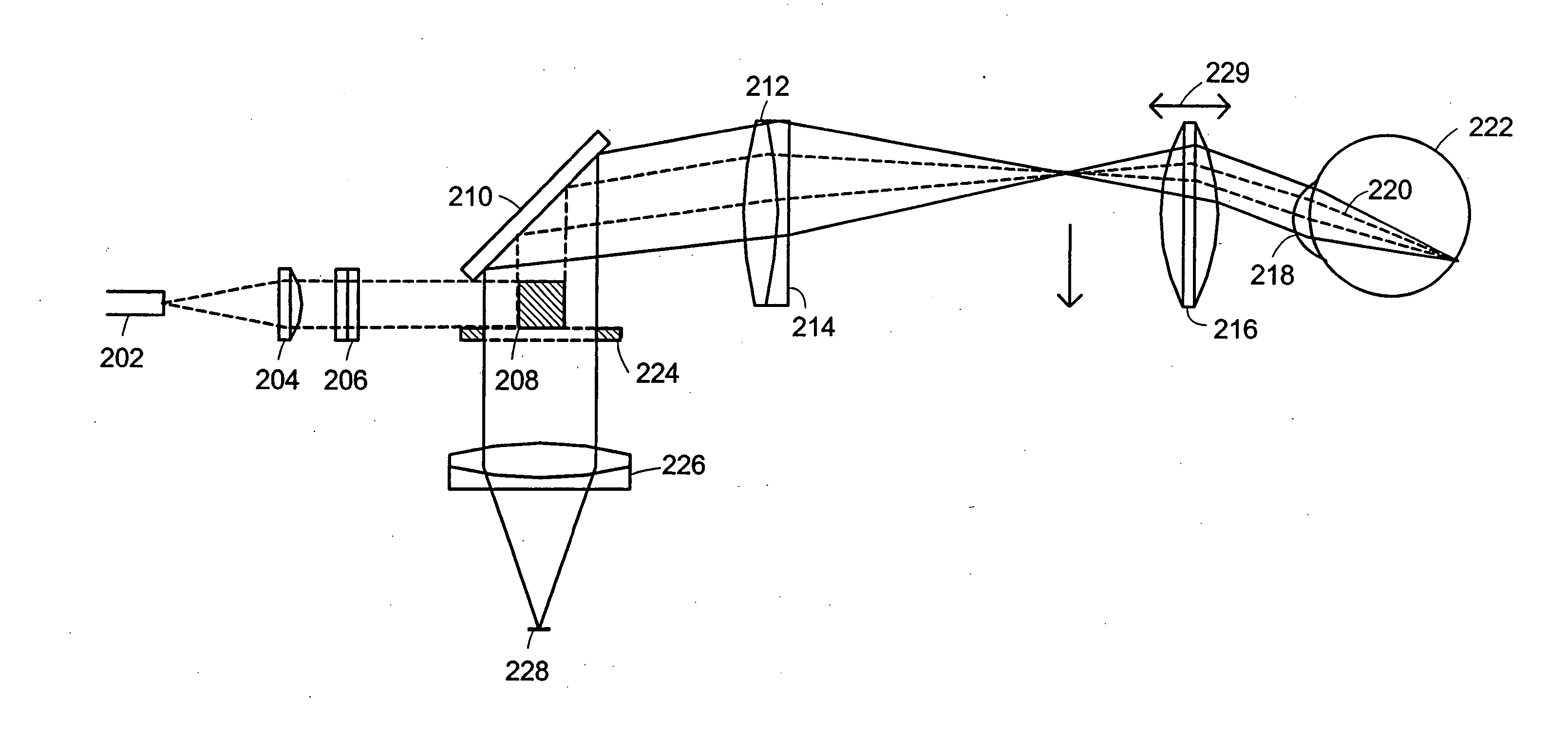
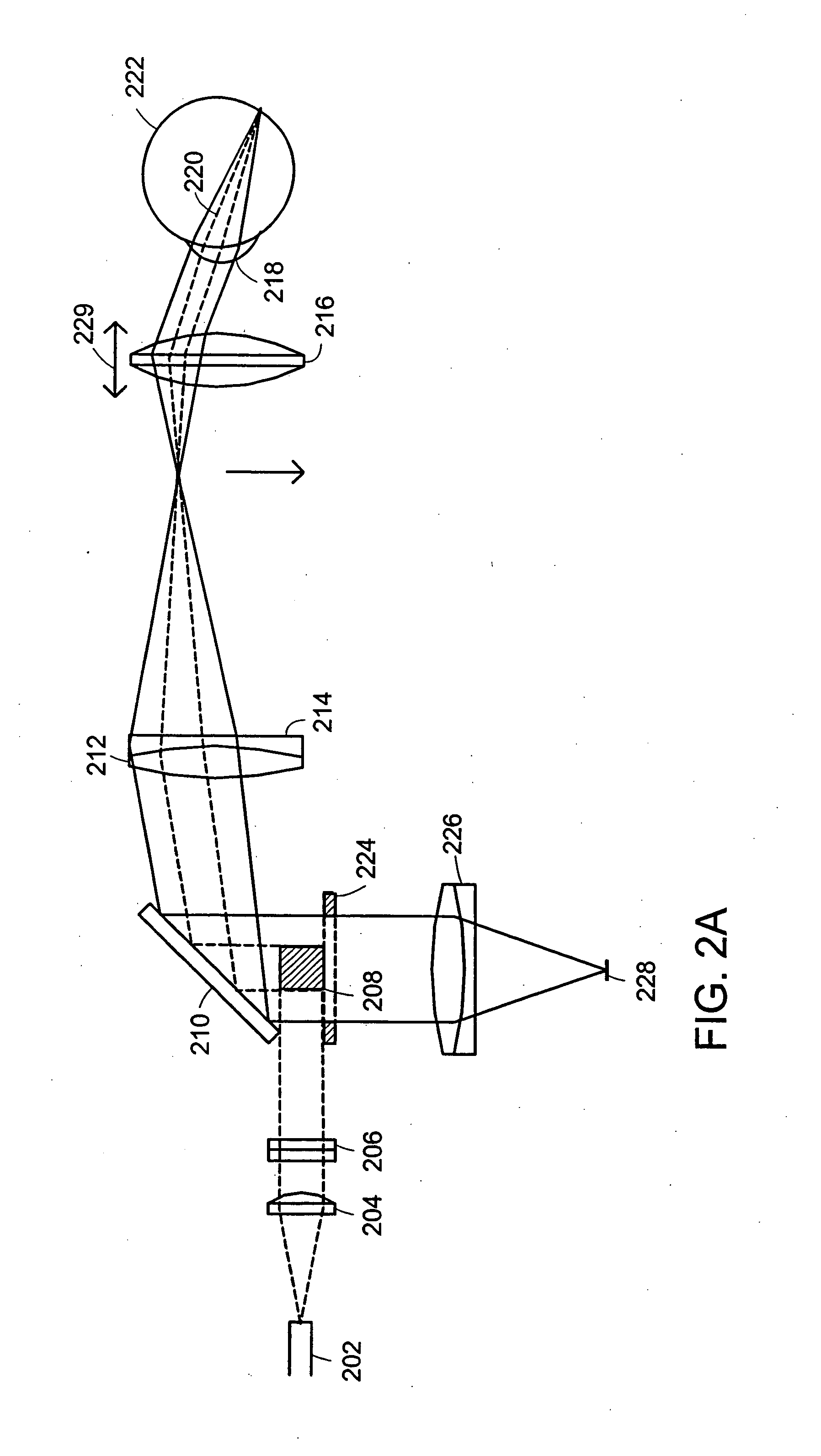
system and method involves a monostatic beam geometry, e.g., the light incoming to the thing to be observed, and the light collected in reflection from the thing, pass through the same location in space between the thing and the optical component nearest the thing. As a result of the monostatic beam geometry, the instrument can be operated with a small, undilated pupil. The instrument remains operative even if the pupil is dilated, however.
There are many benefits that accrue if the pupil of an eye is not required to be dilated for the systems and methods of the invention to function correctly. Dilation is generally performed by applying chemicals topically and waiting for the dilation to occur. The waiting period can be some minutes, typically twenty minutes. Absence of a dilation requirement means that an instrument embodying principles of the invention can be used immediately, rather than only after a delay necessitated by the dilation of the pupil. This allows use in settings such as emergency or field use, where other instruments become useful only after the dilation of the pupil is complete. Dilation of the pupil causes the patient to have reduced visual acuity for periods of up to hours, until the effect of the dilation chemicals wears off. Dilation of the pupil can require a patient to use protective eyewear or to avoid light of ordinary intensity. Dilation of the pupil can cause a patient discomfort. The use of an instrument embodying principles of the invention can eliminate all of the above negative features of dilation of the pupil.
The inventive technology provides an affordable clinical instrument that gives the clinician the power and resolution of the SLO, with some operational features of the most familiar ophthalmic diagnostic instruments, in an untethered package that is comparable in size and weight to commercial hand-held digital video cameras.
The LSLO can provide stereo fundus images. A binocular LSLO, with low-cost wearable display technology and more deeply penetrating near-infrared (NIR) light, can provide real time 3-D morphometric information that is usually the domain of slit-lamp biomicroscopes, binocular indirect ophthalmoscopes (BlOs), and stereo fundus photography at shorter wavelengths. NIR operation increases patient comfort and reduces the risk of phototoxicity during extended exams or procedures. By incorporating additional laser wavelengths as additional channels for particular wavelength combinations, color information can be captured and fused with NIR images. The digital LSLO allows the operator to switch views between live-motion and captured still images with the touch of a button. Synchronous modulation of laser illumination with line-by-line image acquisition and variable scans allows stereo images, dual-color images, or fluorescence images to be multiplexed and recorded. The LSLO can be quickly reconfigured for anterior segment imaging, pupil size and light response. The compact and lightweight LSLO offers the potential for use as a hand-held emergency care aid, particularly with blood in the vitreous from eye or head trauma. A portable digital LSLO which performs some of these functions at a cost approaching indirect ophthalmoscopes, while retaining much of the confocal and NIR advantages of the SLO, becomes more clinically versatile and commercially attractive.
In a preferred embodiment, the scanning mirror that intercepts the redirected line of light and provides a scanned line of light and the scanning mirror that redirects the line of reflected light are the same scanning mirror. In a preferred embodiment, the one or more lenses that focus the scanned line of light on a portion of an eye and the one or more lenses that confocally receive reflected light from the illuminated portion of the eye are the same one or more lenses. In some embodiments, the pupil stop prevents non-confocally received light from proceeding through the optical apparatus.

Problems solved by technology

Dilation of the pupil can cause a patient discomfort.

Method used


Image

  • Line-scan laser ophthalmoscope
  • Line-scan laser ophthalmoscope
  • Line-scan laser ophthalmoscope

Examples


Embodiment Construction

The digital LSLO instrument can be used as a relatively inexpensive multi-mode screening tool to facilitate rapid, non-mydriatic exams for large numbers of patients. In some embodiments of the invention, rapid is to be understood as connoting real time operation. As a portable device, the instrument aids in the early detection of AMD, and other diseases of the elderly, where no economical early warning methods currently exist. The digital LSLO complements existing diagnostics and tele-medicine screening tools for detecting onset of diabetic retinopathy. Many elderly patients may have difficulty in adapting their posture to the demands of any of the standard instruments. Pediatric examination has similar constraints. Instead, instruments should adapt to the needs of the patient. The compact and lightweight LSLO may be used as a hand-held primary care and emergency care aid. The LSLO according to principles of the invention is advantageously used without the necessity to dilate a pup...


PUM


Abstract

Systems and methods for providing a line-scanning laser ophthalmoscope (LSLO) are disclosed. The LSLO uses a substantially point source of light, such as an infrared laser or a super-luminescent diode. The point source is expanded to a line. The LSLO scans the line of light in a direction perpendicular to the line across a region of an eye having an undilated pupil The reflected light is received confocally, using monostatic beam geometry. A beam separator, such as a turning prism or mirror, diverts one of the incoming light and the reflected light to separate the light. An optical stop prevents non-confocally received light from reaching a one-dimensional detector, such as a linear CCD array. An electrical signal responsive to the output light at each of a plurality of locations along the line of output light is processed to provide images of the scanned portion of the eye.

Description

FIELD OF THE INVENTION This invention relates generally to systems and methods for examining eyes. More particularly, the invention relates to systems and methods that employ scanned lines of light for examining eyes. BACKGROUND OF THE INVENTION Fundus imaging is the essential diagnostic procedure in ophthalmology. Instruments of the prior art that are useful for examining the fundus of the eye include direct and indirect ophthalmoscopes, the slit-lamp biomicroscope and the fundus camera. Complementary tools have been developed that broaden diagnostic and therapeutic possibilities, such as the Scanning Laser Ophthalmoscope (SLO). The SLO is a superior tool for rapidly and continuously acquiring high-contrast images of the ocular fundus and its structures, including the distribution of choroidal blood, melanin, and retinal pigments. Because it accommodates a variety of visible and NIR wavelengths, the SLO is especially useful for the study and early diagnosis of diseases such as ag...

Claims


Application Information

Patent Timeline
Patent Type & Authority Applications(United States)
IPC IPC(8): A61B3/12
CPCA61B3/1025
Inventor FERGUSON, R. DANIEL
Owner PHYSICAL SCI
Why Eureka
Try Eureka