Ultrasound coating for enhancing visualization of medical device in ultrasound images

Inactive Publication Date: 2005-04-07
BOSTON SCI SCIMED INC
View PDF41 Cites 109 Cited by
  • Summary
  • Abstract
  • Description
  • Claims
  • Application Information

AI Technical Summary

Benefits of technology

[0008] The invention enhances ultrasonic visualization of a medical device by providing upon the surface of the medical device a contrast agent, preferably comprising microbubbles, that interacts with the illuminating ultrasound differently than does the surrounding ti

Problems solved by technology

However, a limitation of conventional ultrasound imaging is that electrodes are poorly visualized, which inhibits the ability of physicians to properly position the electrodes in the body.
A problem with these techniques is that the surrounding tissue

Method used


Image

  • Ultrasound coating for enhancing visualization of medical device in ultrasound images
  • Ultrasound coating for enhancing visualization of medical device in ultrasound images

Examples


Embodiment Construction

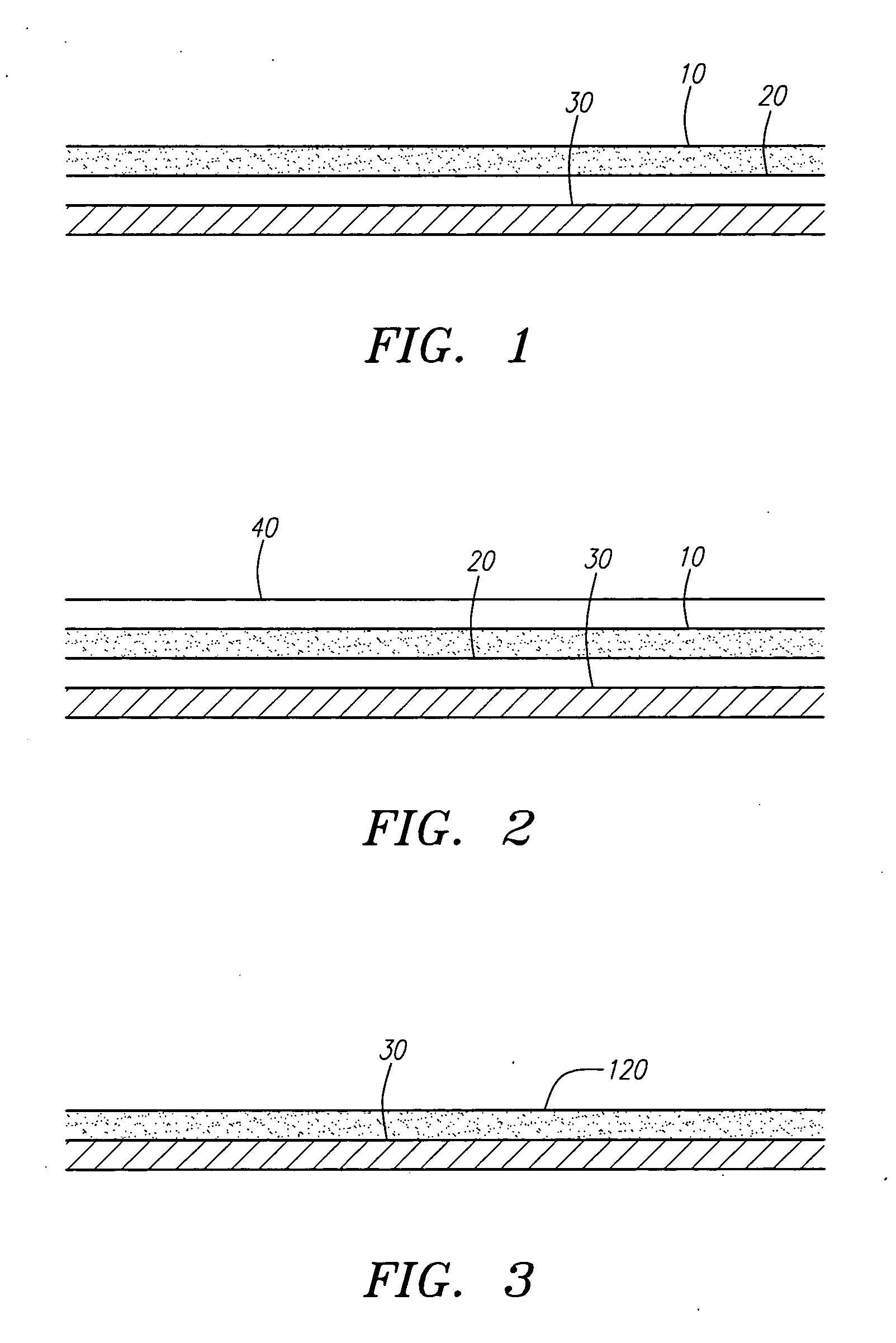
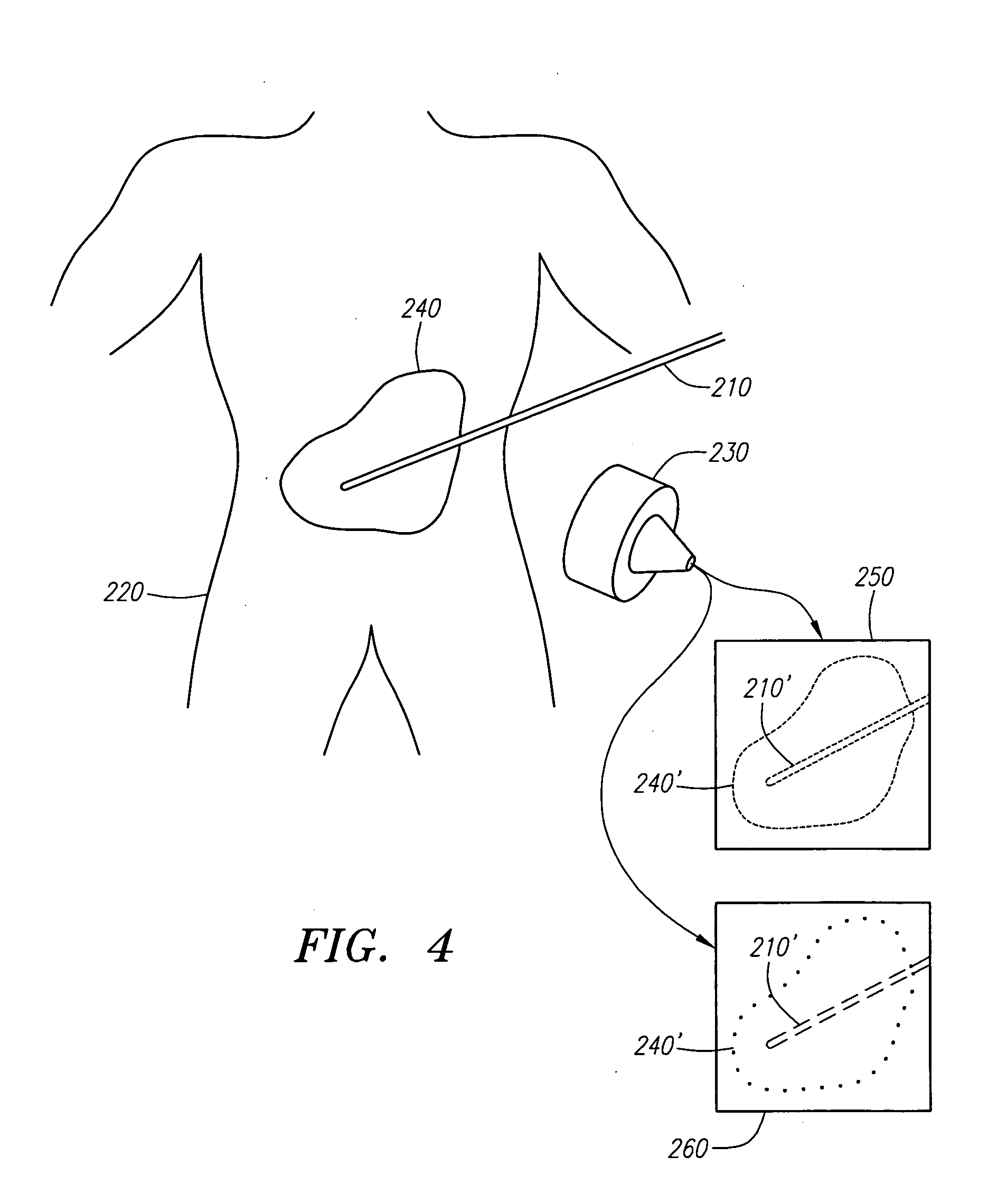
[0019] The invention enhances ultrasonic visualization of a medical device by coating the surface of a medical device with an ultrasound contrast agent. The medical device may be an insertable device, e.g., catheter or guidewire, adapted to perform medical procedures inside the body. The medical device may also be a prosthesis, e.g., stent, adapted to be implanted in the body. The contrast agent should have good contrast properties, i.e., a significant difference in acoustic impedance from the surrounding material.

[0020] In one embodiment, the contrast agents are harmonic ultrasound contrast agents that reradiate incident ultrasound waves nonlinearly and generate vibrations at a harmonic frequency (i.e., a harmonic of the frequency of the incident wave). When used in conjunction with an ultrasound imaging system tuned to the harmonic frequency, the contrast agents enhance visualization of the coated medical device by making the device stand out from the background in the harmonic u...


PUM

PropertyMeasurementUnit
Pressureaaaaaaaaaa
Adhesion strengthaaaaaaaaaa
Sensitivityaaaaaaaaaa

Abstract

The invention enhances ultrasonic visualization of a medical device by coating the surface of the medical device with a contrast agent preferably comprising microbubbles. The contrast agent responds to incident ultrasound waves to generate vibrations at a harmonic frequency (i.e., a harmonic of the frequency of the incident wave), non-harmonic vibrations or a combination of harmonic and non-harmonic vibrations. In one embodiment, the microbubbles are coated on the medical device using an intermediate adhesion layer that adheres to the surface of the device. In a second embodiment, the microbubbles are incorporated in the adhesion layer.

Description

FIELD OF THE INVENTION[0001] The field of the invention relates to contrast agents for ultrasound imaging medical devices, and more particularly, to coating medical devices with ultrasound contrast agents to enhance visualization of the medical devices in ultrasound images. BACKGROUND OF THE INVENTION [0002] Catheters, needles and many other devices are used in minimally-invasive medical procedures to gain access to the interior of the body. The catheters typically include working elements (e.g., electrodes, forceps, snares, angioplasty balloons, stents, and transducers) at or near their distal tip for performing medical procedures within the internal organs and / or orifices of the body for injecting fluids. [0003] In radio frequency (RF) ablation procedures, for example, a catheter equipped with an RF electrode(s) is guided to position the RF electrode at a localized region of the body to be ablated. After the RF electrode is positioned, the RF electrode is energized to emits RF ene...

Claims


Application Information

Patent Timeline
IPC IPC(8): A61K49/22A61L29/08A61L29/18A61L31/08A61L31/10A61L31/18
CPCA61K49/223A61L29/085A61L31/18A61L31/10A61L29/18
Inventor COUVILLON, LUCIEN ALFRED JR.TEO, TAT JIN
Owner BOSTON SCI SCIMED INC
Why Eureka
Try Eureka