Evaluating disease progression using magnetic resonance imaging

a magnetic resonance imaging and disease technology, applied in the field of tracking disease progression using magnetic resonance imaging, can solve the problems of cartilage destruction, severe disability, functional loss of normal matrix physiologic properties, etc., and achieve the effect of accurately determining how and when to treat individual patients, finely targeted treatment research, and effective treatment delivery

Inactive Publication Date: 2005-04-21
PELLETIER JEAN PIERRE +7
View PDF17 Cites 10 Cited by
  • Summary
  • Abstract
  • Description
  • Claims
  • Application Information

AI Technical Summary

Benefits of technology

[0037] Systems and methods according to the invention are advantageous in that they can allow precise quantitative tracking of the progression of diseases, such as rheumatic diseases affecting the cartilage. Such precise quantitative tracking can allow for accurate evaluation of the effects of pharmaceutical agents on these diseases in clinical trials. It may also allow physicians to accurately determine how and when to treat individual patients.
[0038] Systems according to the invention may also provide more insight into disease progression. Because they allow physicians to view the effect of disease on different joint structures, systems according to the invention may permit physicians to gain a more detailed insight into th

Problems solved by technology

Although this disease is often benign, severe degenerative changes may cause serious disability.
Decreased proteoglycan content in conjunction with damaged collagen structure leads to functional loss of normal matrix physiologic properties.
Although the etiology of osteoarthritis is multiple and includes mechanical and biochemical factors, it appears that these culminate in an increased synthesis of proteolytic enzymes by the chondrocytes, which in turn leads to cartilage destruction.
There is no known cure for osteoarthritis, and current treatments are essentially limited to reliving the patient's symptoms.
Unfortunately, evaluating the efficacy of such agents is not an easy, straightforward process.
Simple radiographs are now often used in clinical trials for osteoarthritis to establish inclusion criteria, but such trials have not employed

Method used


Image

  • Evaluating disease progression using magnetic resonance imaging
  • Evaluating disease progression using magnetic resonance imaging
  • Evaluating disease progression using magnetic resonance imaging

Examples


example 1

[0095] Fifteen patients with knee osteoarthritis were recruited from outpatient rheumatology clinics. These patients included male and female individuals satisfying American College of Rheumatologists (ACR) criteria for primary osteoarthritis. They were each symptomatic and required treatment.

[0096] In all cases there was radiological evidence of osteoarthritis in the affected knee, including an X-ray within six months. Each patient exhibited a minimal grade two severity on either space narrowing, osteophyte and / or sclerosis on the Kellgren and Lawrence scale. Absence of chondrocalcinosis was required, and patients with end-stage radiological disease (i.e., grade four) or isolated femoropateilar osteoarthritis were not included in the study.

[0097] Patients were ruled out on the basis of a number of possibly confounding conditions, including secondary osteoarthrits, inflammatory arthritis, post-traumatic arthritis, metabolic arthritis, septic arthritis, crystal-induced disease, Pag...

example 2

[0102] Thirty-five patients with knee osteoarthritis were recruited from outpatient rheumatology clinics using similar criteria to those used for the first Example. The patients exhibited the baseline demographics presented in Table 1.

TABLE 1Age (yrs.) 63.1 (9.1)Womac Pain59.4 (3.93)% female  74%Womac Stiff.45.7 (4.77)Weight (kg) 84.1 (15.1)Womac Fnct.60.3 (3.99)% Analg.82.6%Womac Total56.9 (3.99)% NSAIDs  77%Patient Global54.5 (3.74)SF-36 PCS37.1 (1.65)50 Walk (sec) 11.6 (3.6)VAS PAIN48.2 (4.97)ROM (deg.)126.9 (12.2)MD Global59.8 (3.12)

(VAS scores 100 = worst)

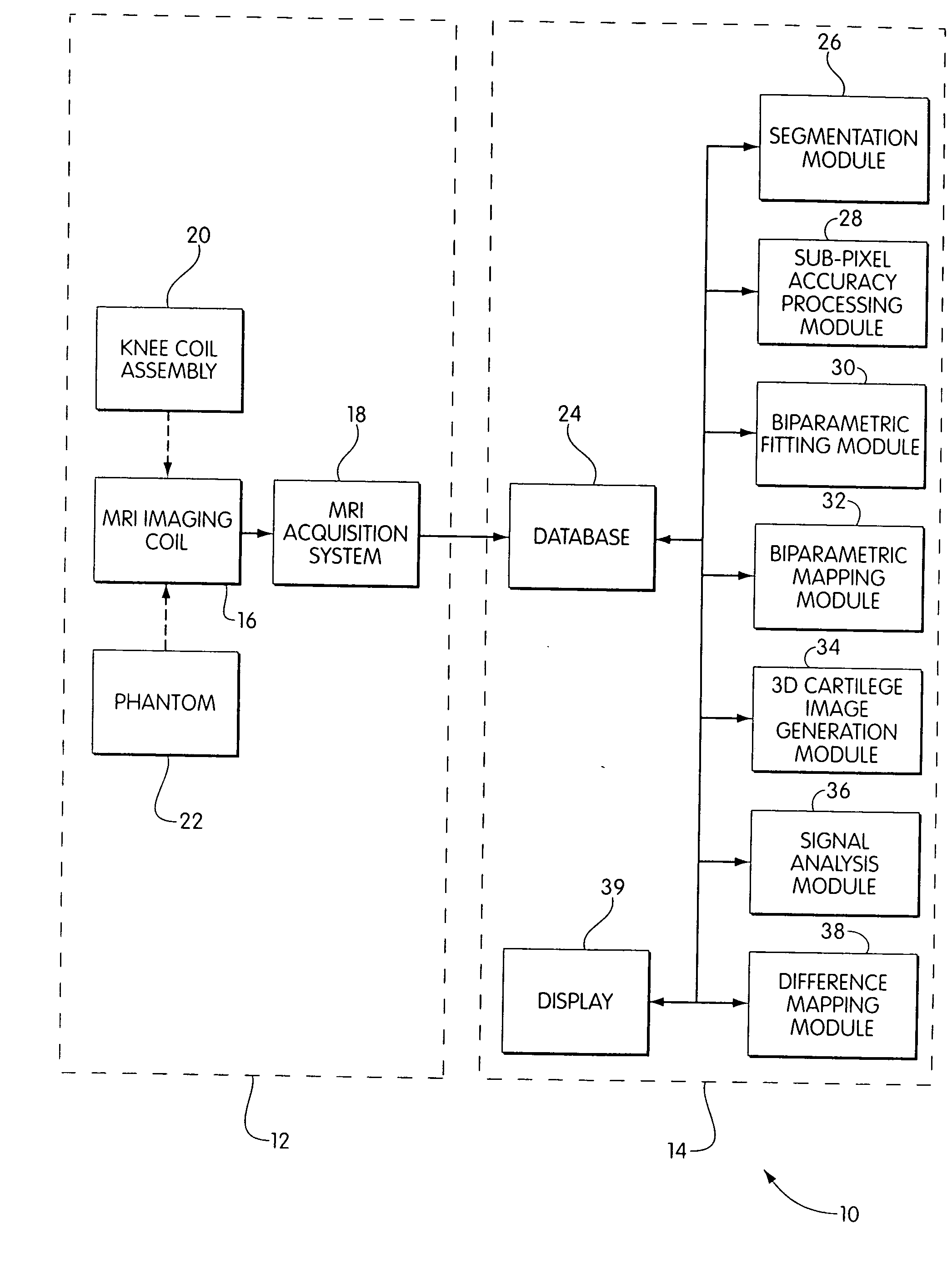
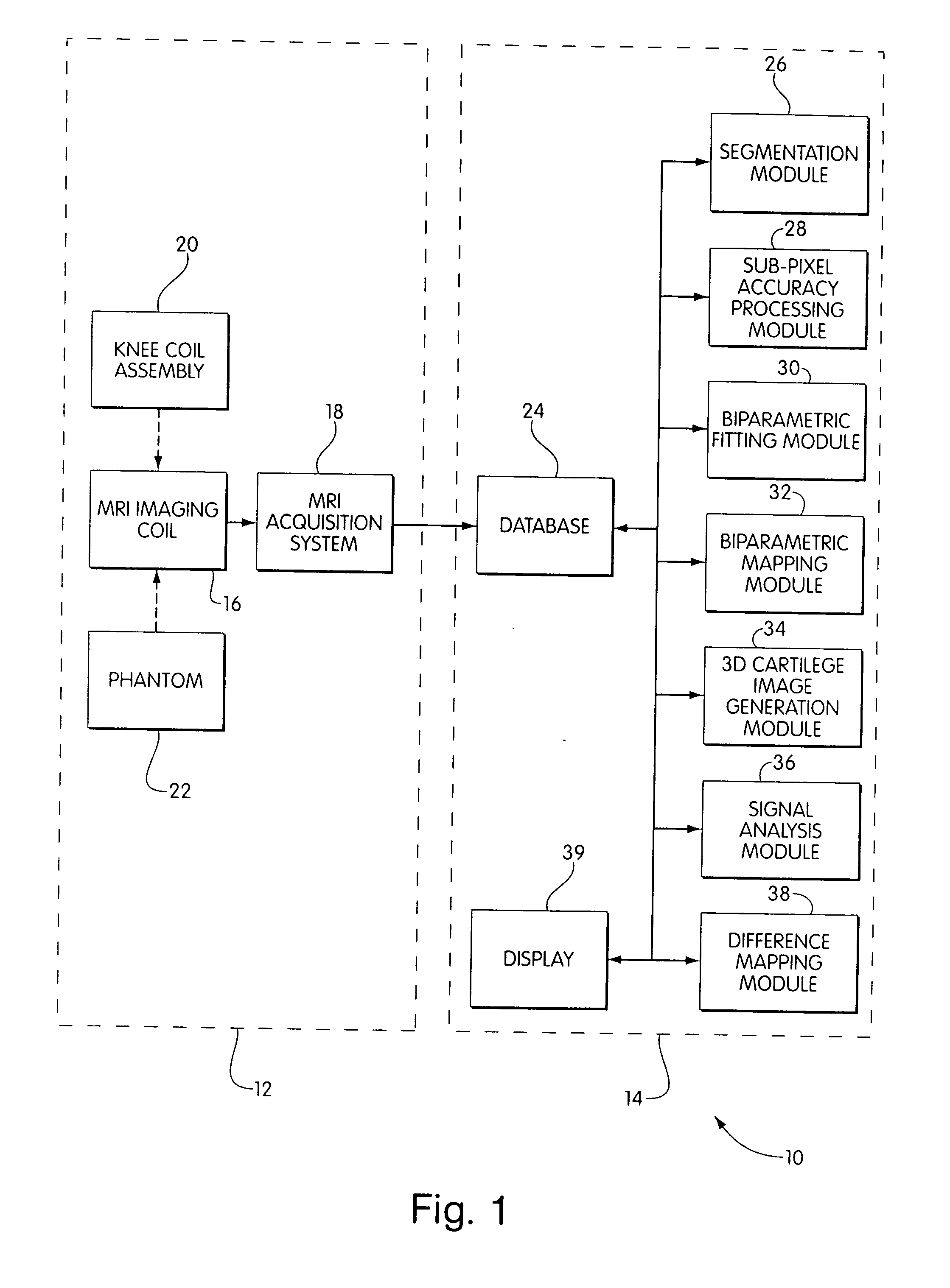
[0103] The patients were assessed at baseline, six months, and one year using an MRI system generally comparable to that described above. As part of this assessment, the images obtained were systematically analyzed and quantified using a processing system generally comparable to that described above. Imaging parameters were: Voxel size: 0.3×0.4×1 mm, with a 512×410 grid; 3D FISP; TR=42, and TE=7.

[0104] The total cartilage ...


PUM


Abstract

An orthopedic magnetic resonance imaging system is disclosed. This system includes a source of magnetic resonance imaging data sets resulting from successive magnetic resonance imaging acquisitions from a diseased joint of a patient. A segmentation module segments surfaces in the joint based on information contained within at least one of the data sets, and a registration module spatially registers, in three dimensions, information represented by a first of the data sets with respect to information represented by one or more further data sets for the same patient. A comparison module detects differences between information represented by the data sets caused by progression of the disease in the joint of the patient between acquisitions. A cross-patient comparison module can compare detected differences for the patient with detected differences for at least one other patient.

Description

CROSS-REFERENCE TO RELATED APPLICATION [0001] This application is a continuation of U.S. Ser. No. 10 / 430,800, filed May 6, 2003, which is a continuation of U.S. Ser. No. 09 / 704,269, filed Nov. 1, 2000, which claims the benefit of U.S. Provisional Application no. 60 / 162,871, filed Nov. 1, 1999, all of which are herein incorporated by reference.FIELD OF THE INVENTION [0002] This invention relates to methods and apparatus for tracking disease progression using magnetic resonance imaging, including methods and apparatus for efficiently and precisely tracking the progression of rheumatic diseases affecting cartilage. BACKGROUND OF THE INVENTION [0003] Osteoarthritis is a prevalent disease characterized mainly by cartilage degradation that is clinically reflected by a gradual development of joint pain, stiffness, and loss of motion. Osteoarthritis is extremely frequent in the general population, and it is estimated that its radiological prevalence is close to 50% overall. This figure is e...

Claims


Application Information

Patent Timeline
IPC IPC(8): G01R33/56G06T5/50G06T7/00
CPCA61B5/4504A61B5/4514A61B5/4528G06T7/0012G01R33/5608G06T5/50G01R33/56
Inventor PELLETIER, JEAN PIERREPELLETIER, JOHANEGUISE, JACQUES DERAYNAULD, JEAN-PIERREBARTHIAUME, MARIE-JOSEEBEAUDOIN, GILLESGODBOUT, BENOITKAUFFMANN, CLAUDE
Owner PELLETIER JEAN PIERRE
Why Eureka
Try Eureka