Process to study changes in gene expression in granulocytic cells

a technology of granulocytic cells and gene expression, applied in the field of process to study changes in gene expression in granulocytic cells, can solve the problems of eosinophil cationic, systematic approach to assess transcriptional response, increased blood flow (hyperemia) and edema

Inactive Publication Date: 2005-04-21
GENE LOGIC +2
View PDF0 Cites 6 Cited by
  • Summary
  • Abstract
  • Description
  • Claims
  • Application Information

AI Technical Summary

Problems solved by technology

Vasoconstriction is followed within seconds by the acute vascular response resulting in increased blood flow (hyperemia) and edema.
No study, however, has taken a systematic approach to assess the transcriptional response during neutrophil activation via contact with a pathogen or from neutrophils isolated from a subject with a sterile inflammatory disease.
Eosinophil cationic proteins are highly toxic for schistosomes and may be responsible for binding of eosinophils to parasitic worms as well as fragmentation of the parasite.

Method used


Image

  • Process to study changes in gene expression in granulocytic cells
  • Process to study changes in gene expression in granulocytic cells
  • Process to study changes in gene expression in granulocytic cells

Examples


example 1

Production of Gene Expression Profiles Generated from cDNAs Made with RNA Isolated from Neutrophils Exposed to Virulent and Avirulent Bacteria.

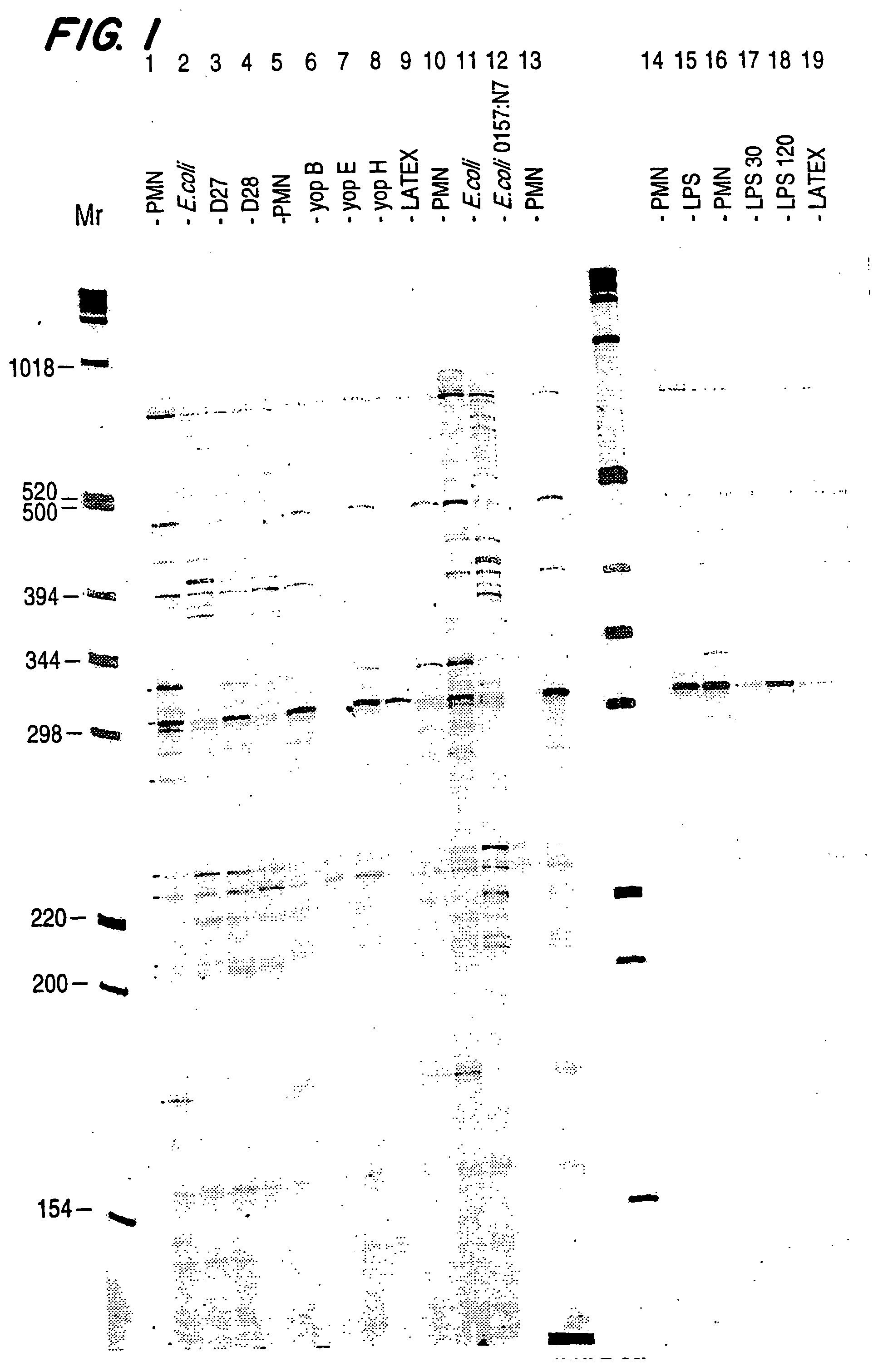
[0078] Expression profiles of RNA expression levels from neutrophils exposed to various bacteria offer a powerful means of identifying genes that are specifically regulated in response to bacterial infection. As an example, the production of expression profiles from neutrophils exposed to virulent and avirulent E. coli and Y. pestis allow the identification of neutrophil genes that are specifically regulated in response to bacterial infection.

[0079] Neutrophils were isolated from normal donor peripheral blood following the LPS-free method. Peripheral blood was isolated using a butterfly needle and a syringe containing 5 cc ACD, 5 cc of 6% Dextran (in normal saline). After 30 minutes of settling, plasma was collected and HBSS (without Ca++ or Mg++) was added to a total volume of 40 ml. The plasma was centrifuged (1500 rpm, for 15 m at 4° C....

example 2

Production of Gene Expression Profiles Generated from cDNAs Made with RNA Isolated from Neutrophils Exposed to Virulent and Avirulent Bacteria and Neutrophils Exposed to Cytokines.

[0088] Neutrophils were isolated from normal donor peripheral blood following the LPS-free method as set forth in Example 1.

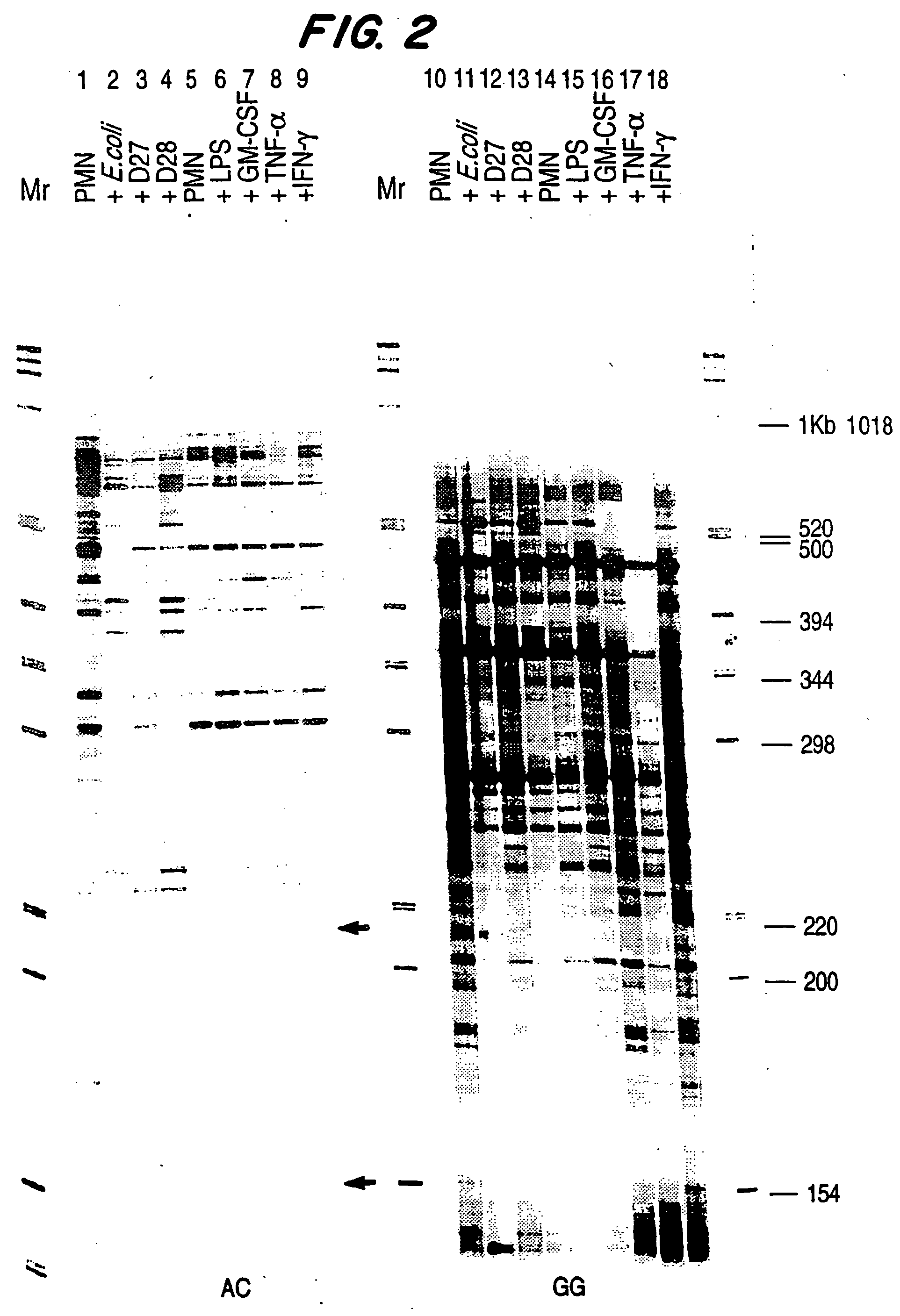
[0089] Neutrophils were incubated with virulent and avirulent E. coli or Y. pestis, LPS at 1 ng / ml, GM-CSF at 100 units / ml, TNFα at 1000 units / ml, or γIFN at 100 units / ml. The bacterial cells, LPS or cytokines were added to approximately 3.38×10⁸ cells in 100 ml of RPMI containing 6% H1 autologous serum. Incubation proceeded for 2 to 4 hours, preferably 2 hours, with gentle rotation in disposable polycarbonate Erlenmeyer flasks at 37° C. After incubation, the cells were spun down and washed once with HBSS.

[0090] After incubation of the neutrophils, RNA was extracted and the cDNA profiles prepared as described in Example 1. FIG. 2 is an autoradiogram of the expression profiles gene...

example 3

Production of Gene Expression Profiles Generated from cDNAs Made with RNA Isolated from Neutrophils Exposed to Bacteria Using all 12 Possible Anchoring Oligo d(T) n1,n2.

[0092] Neutrophils were isolated from normal donor peripheral blood following the LPS-free method.

[0093] Neutrophils were incubated with E. coli or Y. pestis.

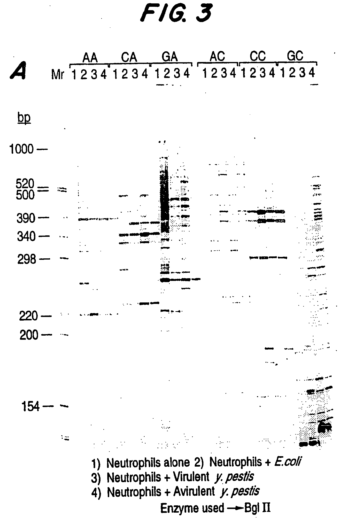
[0094] After incubation of the neutrophils, RNA was extracted and the cDNA profiles prepared as described in Example 1. FIG. 3 is an autoradiogram of the expression profiles generated from cDNAs made with RNA isolated from control (untreated) neutrophils (lane 1), neutrophils incubated with avirulent E. coli K12 (lane 2), virulent Y. pestis (lane 3), avirulent Y. pestis (lane 4). The anchoring oligo d(T)18 n1 and n2 positions are indicated at the top of the figure. The cDNAs were digested with BglII.

[0095]FIG. 4 represents a summary of genes which are differentially expressed in neutrophils upon exposure to virulent and avirulent E. coli and Y. pestis. Expr...


PUM

PropertyMeasurementUnit
total volumeaaaaaaaaaa
concentrationaaaaaaaaaa

Abstract

The present invention comprises a method to identify granulocytic cell genes that are differentially expressed upon exposure to a pathogen or in a sterile inflammatory disease by preparing a gene expression profile of a granulocytic cell population exposed to a pathogen or isolated from a subject having a sterile inflammatory disease and comparing that profile to a profile prepared from quiescent granulocytic cells. The present invention is particularly useful for identifying cytokine genes, genes encoding cell surface receptors and genes encoding intermediary signaling molecules. The invention also includes methods to identify a therapeutic agent that modulates the expression of at least one gene in a granulocyte population. Genes which are differentially expressed during neutrophil contact with a pathogen, such as a virulent bacteria, or that are differentially expressed in a subject having a sterile inflammatory disease are of particular importance.

Description

[0001] This application is related to application Ser. No. 08 / 510,032, Ser. No. 60 / 056,844 and application Ser. No. 08 / 688,514, all of which are herein incorporated by reference in their entirety. All published articles, patents and other publications cited throughout this application are herein incorporated by reference in their entirety.TECHNICAL FIELD [0002] This invention relates to compositions and methods useful to identify agents that modulate the response of granulocytes to inflammatory and infectious conditions. BACKGROUND OF THE INVENTION [0003] Granulocytes (i.e., neutrophils, eosinophils and basophils) are involved in the immune response elicited by inflammation and infection. Inflammation [0004] Inflammation is a localized protective response elicited by injury or destruction of tissues which serves to destroy, dilute or wall off both the injurious agent and the injured tissue. It is characterized by fenestration of the microvasculature, leakages of the elements of blo...

Claims


Application Information

Patent Timeline
Patent Type & Authority Applications(United States)
IPC IPC(8): C12Q1/68C12Q1/6809C12Q1/6883
CPCC12Q1/6809C12Q2600/158C12Q2600/142C12Q1/6883
Inventor YERRAMILLI, SUBRAHMANYAMPRASHAR, YATINDRANEWBURGER, PETERGOGUEN, JONWEISSMAN, SHERMAN
Owner GENE LOGIC
Why Eureka
Try Eureka