Compositions and methods for enhancing phagocytosis or phagocyte activity

a phagocytosis and phagocyte technology, applied in the direction of liposomal delivery, depsipeptides, peptide/protein ingredients, etc., can solve the problems of insufficient immune surveillance to prevent cancer, insufficient body's own mechanisms for detecting and removing cells, and often insufficient harmful noncellular molecular entities, etc., to achieve better targets for phagocytosis, enhance or incite phagocytosis

Active Publication Date: 2005-05-26
APELLIS PHARMA
View PDF5 Cites 140 Cited by
  • Summary
  • Abstract
  • Description
  • Claims
  • Application Information

AI Technical Summary

Benefits of technology

[0004] The present invention addresses the foregoing needs, among others. The invention provides a system for tagging cells or noncellular molecular entities that may occur in or on biological systems such as living cells, tissues, organs, organisms, body fluids, etc., with molecules that increase the likelihood that a target cell or noncellular molecular entity will be phagocytosed, e.g., that make the tagged cells or noncellular molecular entities better targets for phagocytosis. In particular, the present invention provides the recognition that cells can be tagged or coated with molecules that incit

Problems solved by technology

The body has a number of mechanisms by which to rid itself of undesirable cells, and many diseases result from defects or deficiencies in these processes.
For example, although the immune system is believed to play an important role in preventing the development of tumors or curtailing their growth or spread, it is clear that in many cases immune surveillance is not sufficient to prevent cancer.
Proliferation of vascular tissue in the eye can lead to vision loss.
Molecules and other entities that are not properly removed or disposed of can also cause disease.
Thus it i

Method used


Image

  • Compositions and methods for enhancing phagocytosis or phagocyte activity
  • Compositions and methods for enhancing phagocytosis or phagocyte activity
  • Compositions and methods for enhancing phagocytosis or phagocyte activity

Examples


example 1

Enhancement of Phagocytosis by a PS Derivative

[0290] Materials and Methods

[0291] Cells and Cell Culture.

[0292] Human umbilical vein endothelial cells (HUVECs) and MonoMac-1 cells (a human myeloma cell line) were obtained from Cambrex Inc., East Rutherford, N.J., and were maintained under standard culture conditions. Both cell lines are adherent under these culture conditions.

[0293] Compound Synthesis.

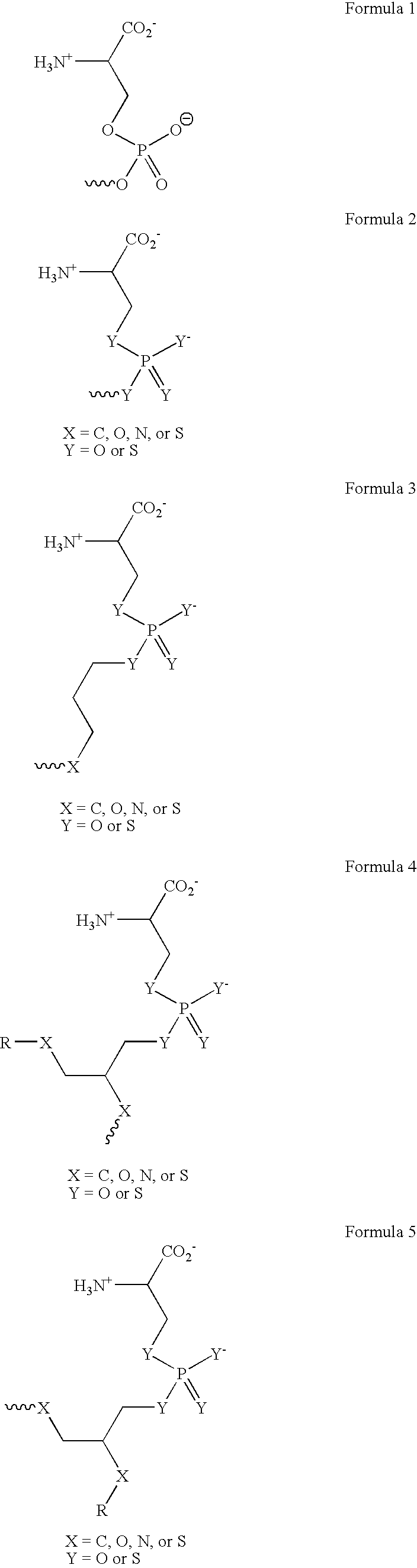
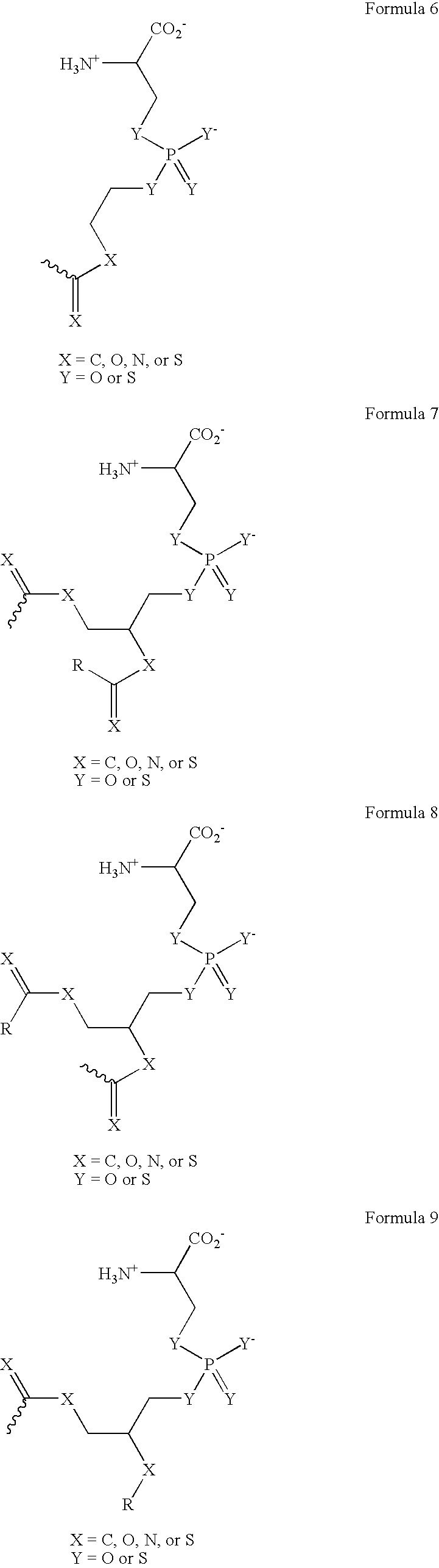
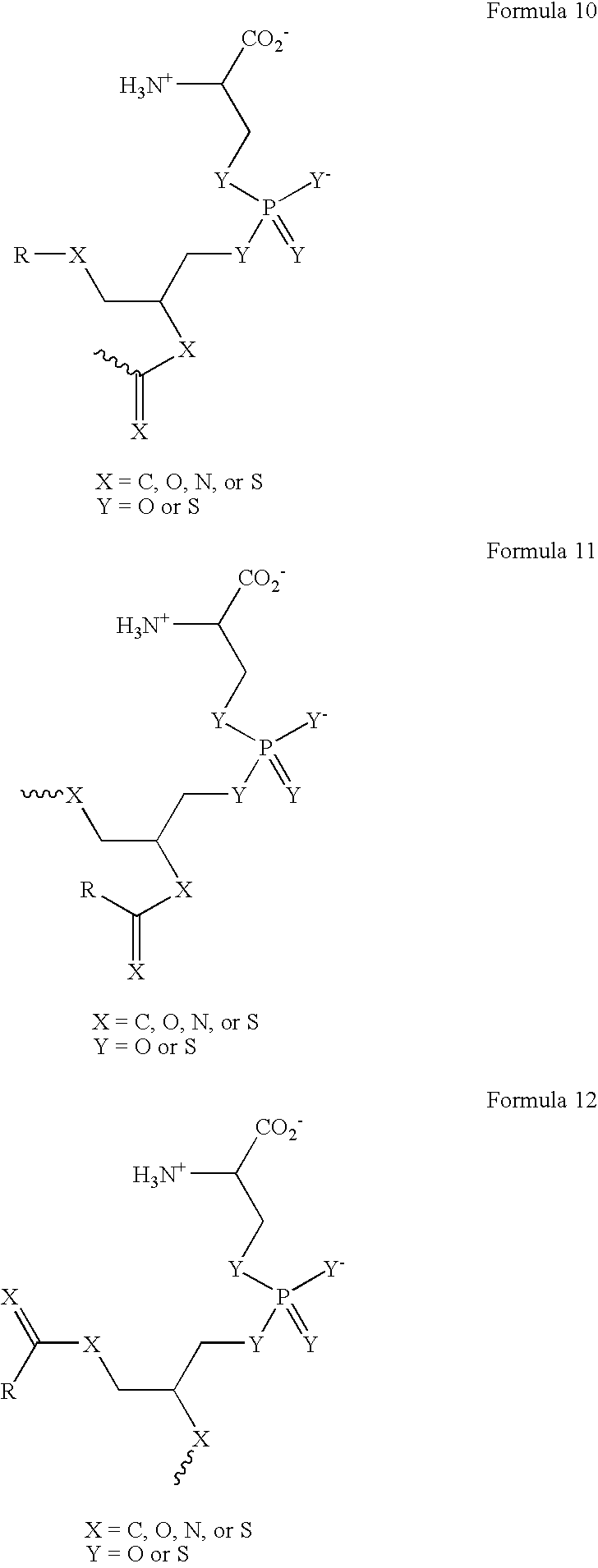
[0294] The phosphatidylserine derivative shown in FIG. 4C, containing a PS headgroup, was synthesized according to the scheme shown in FIG. 4A, which is described above. The commercially available phosphatidylcholine derivative 1-Hexanoyl-2-Hydroxy-sn-Glycero-3-Phosphocholine (Cat. No. 855175 Avanti Polar Lipids, Alabaster, Ala.) was used, resulting in a compound in which R is a 5 carbon chain. The PS derivative reacts with free thiols in proteins on the cell surface. In other experiments, the phosphatidylcholine derivative 1-Myristoyl-2-Hydroxy-sn-Glycero-3-Phosphocholine or 1-Tet...

example 2

Synthesis of an MGF-E8-Streptavidin Fusion Protein

[0308] MFG-E8 (GenBank: NM—0045928) is a protein of 387 amino acids. Two splicing variants of this protein exist, an S form and an L form. While they differ only by a proline-rich motif present in the L form, the L form has been demonstrated to be more active and is used here.

[0309] Obtaining MFG-E8 cDNA. Human MFG-E8 is obtained by PCR from a first-strand cDNA library obtained from human fetal spleen tissue (BD Clonetics). An epitope tag (FLAG) is then incorporated into the N-terminal part of the protein between the signal peptide (amino acid residues 1-22) and the first EGF-like domain (amino acid residues 24-61). This tag can be used for monitoring protein expression by Western analysis and for purification with the use of anti-FLAG antibodies and anti-FLAG M2 affinity gel (Sigma) respectively.

[0310] Fusing MFG-E8 with SAV. Fusing MFG-E8 to SAV (GenBank: X03591) should provide MFG-E8 with the ability to bind cells that have bee...

example 3

Effect of MFG-E8 Derivatives on Phagocytosis

[0314] This example describes assessment of the ability of two different MFG-E8 derivatives to enhance phagocytosis of target cells in vitro. The target cells used here are endothelial precursor cells (EPCs) (101). These cells exhibit a gene expression profile more similar to that of cells isolated from fresh surgical specimens of human tumors than HUVECs and may thus be preferable for use in such assays. In addition, the phagocytic cells are human blood-derived (HBD) macrophages rather than MonoMac-1 s.

[0315] Preparation of EPC and macrophage cell cultures. EPCs are prepared as described by Bagley et al (101). CD34+ / AC133+ progenitor cells from human bone marrow cells will be purchased from Cambrex. They will be grown at 1-2×10⁵ cells / ml in IMDM medium (Cambrex) supplemented with 15% FBS (Invitrogen Corp., Carlsbad, Calif.), 50 ng / ml VEGF165 (R&D Systems, Minneapolis, Minn.), 50 ng / ml rhbFGF (R&D Systems), and 5 units / ml heparin (Sigma,...


PUM

PropertyMeasurementUnit
Timeaaaaaaaaaa
Compositionaaaaaaaaaa
Structureaaaaaaaaaa

Abstract

The present invention provides a system for enhancing clearance or destruction of undesirable cells or noncellular molecular entities by tagging such cells or noncellular molecular entities with a marker that targets the cells or noncellular molecular entities for phagocytosis (phagocytic marker). The target cells can be, for example, endothelial cells, tumor cells, leukocytes, or virus-infected cells. In certain embodiments of the invention the tagging is accomplished by administering a composition comprising an antibody or ligand linked to the phagcytotic marker, wherein the antibody or ligand binds to a cell type specific marker present on or in the cell surface of a target cell. In preferred embodiments of the invention, the phagocytic marker comprises phosphatidylserine or a group derived from phosphatidylserine, thrombospondin-1, annexin I, or a derivative of any of these.

Description

CROSS-REFERENCE TO RELATED APPLICATIONS [0001] This application claims priority to provisional application 60 / 497,086, filed Aug. 22, 2003; 60 / 514,941, filed Oct. 28, 2003; 60 / 523,611, filed Nov. 19, 2003; 60 / 524,126, filed Nov. 21, 2003; 60 / 524,730, filed Nov. 24, 2003, and 60 / 547,951, filed Feb. 26, 2004, all of which are herein incorporated by reference.BACKGROUND OF THE INVENTION [0002] The body has a number of mechanisms by which to rid itself of undesirable cells, and many diseases result from defects or deficiencies in these processes. For example, although the immune system is believed to play an important role in preventing the development of tumors or curtailing their growth or spread, it is clear that in many cases immune surveillance is not sufficient to prevent cancer. Particular populations of cells can also cause or contribute to a variety of other diseases. For example, white blood cells of various types may cause rejection of a transplanted organ, auto-immune diseas...

Claims


Application Information

Patent Timeline
IPC IPC(8): A61K38/17A61K47/48C12N5/08
CPCA61K47/48053A61K47/48238B82Y5/00A61K47/48823A61K47/48815A61K47/544A61K47/62A61K47/6911A61K47/6913A61P17/02A61P19/10A61P3/04A61P35/00A61P37/00A61P7/02A61P9/10
Inventor FRANCOIS, CEDRICDESCHATELETS, PASCALOLSON, PAULMACHIELS, ALEC
Owner APELLIS PHARMA
Why Eureka
Try Eureka