
Application of the kelvin probe technique to mammalian skin and other epithelial structures

a technology of kelvin probe and mammalian skin, applied in the field of application of kelvin probe to mammalian skin and other epithelial structures, can solve the problems of increasing the risk of procedure, no consistent methodology established for the use of electric fields in the treatment of wounds, and limited standard techniques for determining this information

Inactive Publication Date: 2005-07-14
NUCCITELLI RICHARD +2
View PDF8 Cites 11 Cited by
  • Summary
  • Abstract
  • Description
  • Claims
  • Application Information

AI Technical Summary

Benefits of technology

[0021] One aspect of the present invention is directed to a probe that will measure the electric fields in and surrounding a wound or skin lesion in a mammal. The probe is comprised of a vibrating metallic probe tip that is placed close to the skin in the air. This eliminates the need for penetrating electrodes or contact electrodes and another advantage is that electromagnetic shielding is unnecessary.

Problems solved by technology

However, there has been no consistent methodology established for the use of electric fields in the treatment of wounds.
This inconsistency is due to the lack of reliable information regarding the electric fields associated with normally healing wounds in humans.
Standard techniques for determining this information are limited.
Both the glass microelectrodes and the chlorided silver wire surfaces are fragile and either break (glass) or are damaged (AgCl) during skin puncture, increasing the risk of the procedure.
Electrode tip placement, both the depth and relative lateral spacing, is difficult to reliably reproduce because the electrodes must be positioned using a micromanipulator that is mounted on a support that is usually not directly attached to the subject under study.
In addition, to reduce noise, the measurement set-up must be placed in an electromagnetically shielded cage, which would severely hamper the portability and ultimate patient utility of the measurement system.
The use of surface skin electrodes also presents problems.
Due to the variability of the resistance of the stratum corneum from day to day, body location and emotional state, it is difficult to reliably measure very small, potential variations (several millivolts) over small distances on the order of 100 μm.
However, this method has several problems when applied to mammals.
While anesthesia can be used to immobilize a subject, this poses unacceptable health risks to humans.
This constant movement poses a serious problem for making accurate measurements.
Sources of artifactual signals in mammalian skin make accurate skin surface potential measurements difficult.

Method used


Image

  • Application of the kelvin probe technique to mammalian skin and other epithelial structures
  • Application of the kelvin probe technique to mammalian skin and other epithelial structures
  • Application of the kelvin probe technique to mammalian skin and other epithelial structures

Examples


Embodiment Construction

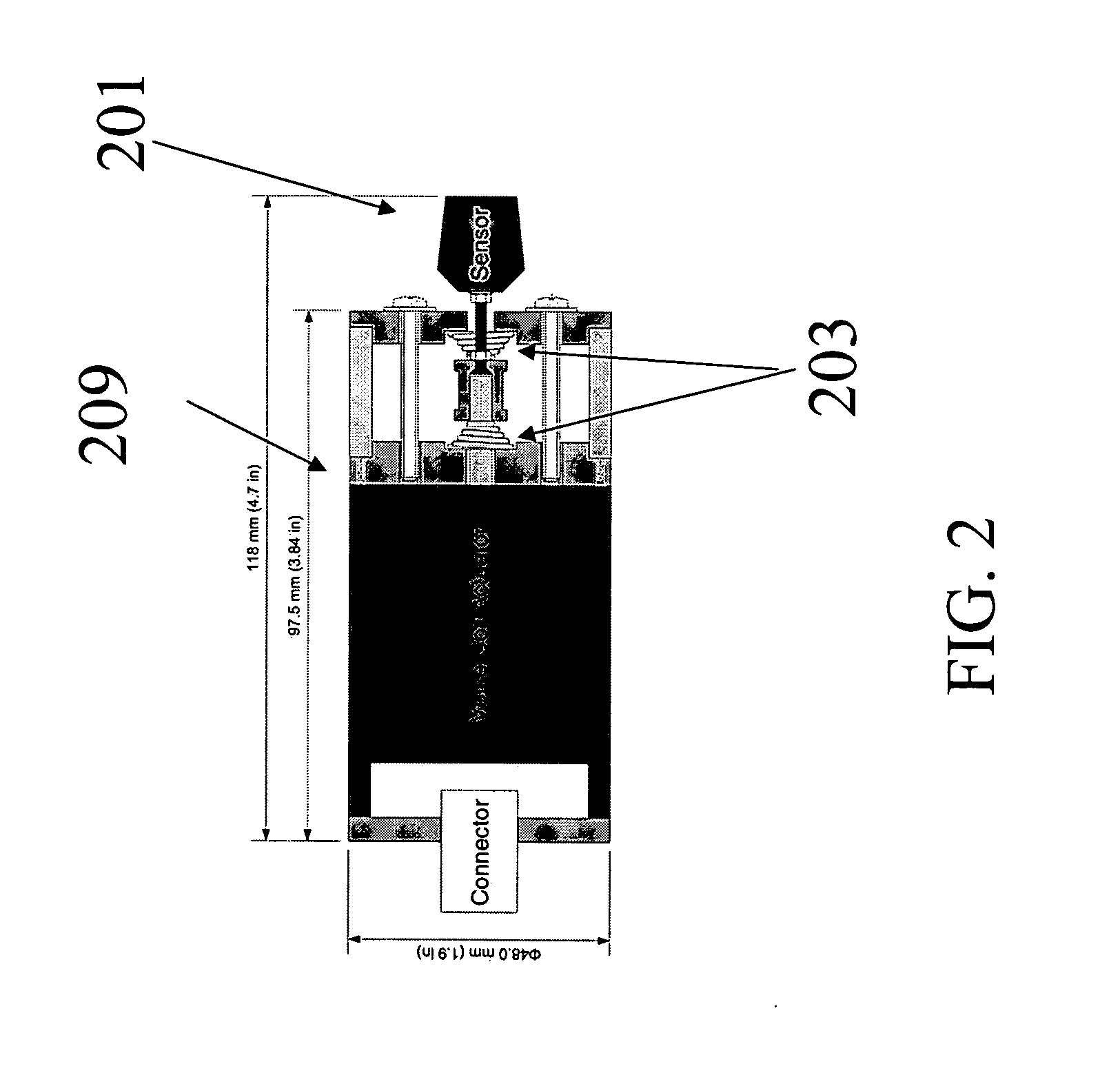
[0048]FIG. 2 depicts the probe vibrator and head that is used in one embodiment of the present invention. Probe head 201 is comprised of one or many small metal plates that each have a surface area of 0.2 mm2, but a different size can be used and still be within the scope of the present invention. Each plate is connected to a low noise current-to-voltage (I / V) converter housed within the probe head 201 and a copper shield held at the reference potential surrounds the entire head. The vibrator unit 209 is designed to move the probe head along a single axis that is perpendicular to the surface of the skin or epithelia under study. This can be achieved with many types of vibrators based on piezoelectric, magnetostrictive or electromagnetic transducers. One embodiment of the present invention is composed of an cylinder that is 1.9″ in diameter and 4.7″ long and contains an electromagnetic transducer or voice coil actuator, commercially available from BEI Kimco. This suspension system us...


PUM


Abstract

A system and method is disclosed for obtaining measurements of the electric fields around skin wounds and lesions on mammals noninvasively. The system and method is comprised of a vibrating metallic probe tip that is placed close to the skin in the air. By applying a series of known voltages to the metal probe tip or to the skin beneath it, the skin's local surface potential can be measured and the lateral electric field can be calculated from the spatial distribution of surface potential measurements. Surface artifacts that can affect the measurements are removed and active feedback is used to maintain a constant distance between the probe and the skin surface.

Description

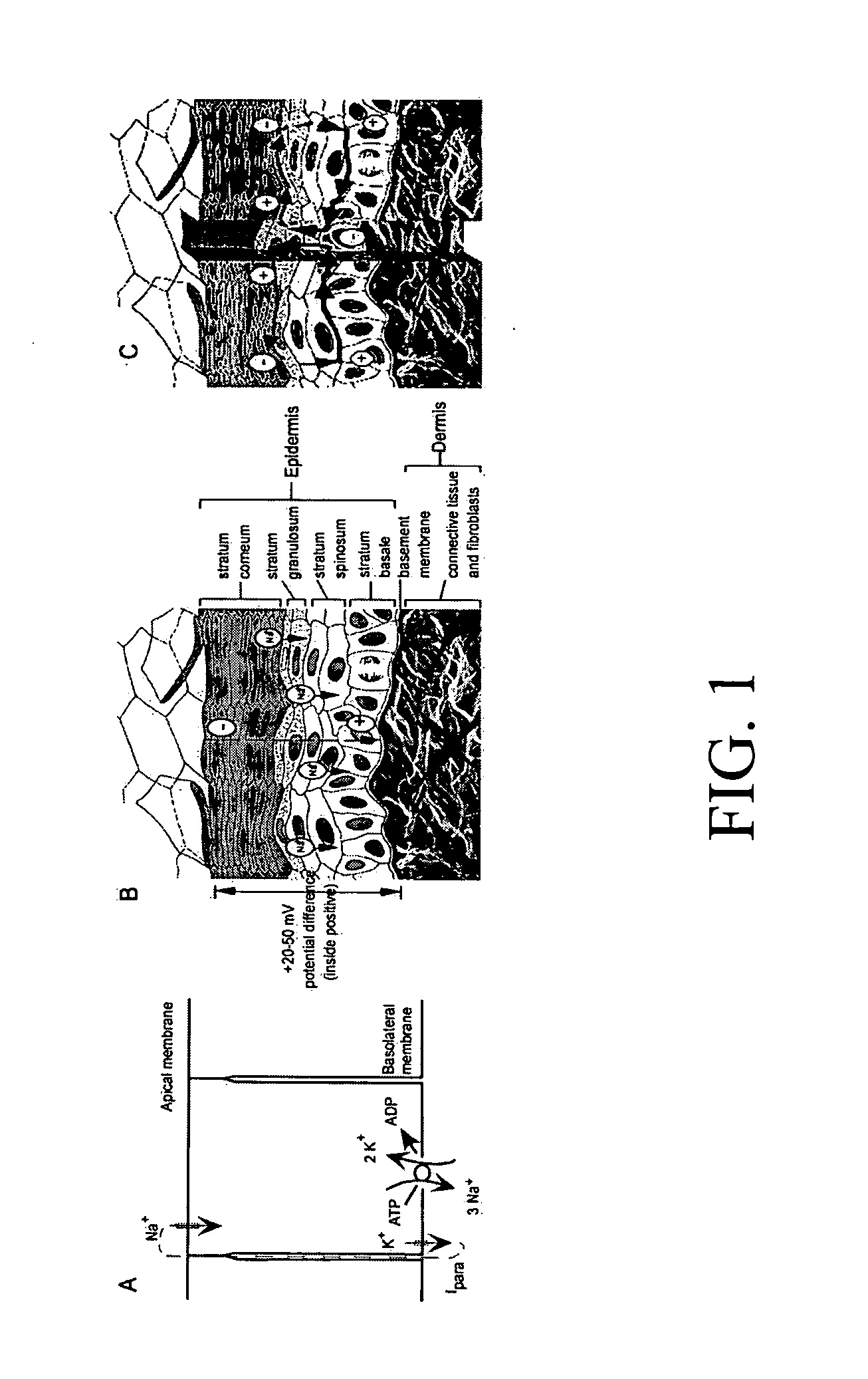
CROSS-REFERENCE TO RELATED APPLICATION [0001] This application claims the benefits of U.S. provisional application Ser. No. 60 / 534,910, filed Jan. 8, 2004, which is incorporated herein by reference in its entirety.FIELD OF THE INVENTION [0002] This application is directed to a method and system for acquiring information from skin and other epithelia. More particularly, this application is directed to the use of this information as a diagnostic tool. BACKGROUND OF THE INVENTION [0003] It is known that ionic currents exit skin wounds. The ultimate driving force for all wound currents is the voltage generated across the epidermis. The epidermis of the skin normally generates a voltage across itself, termed the transepithelial potential, by pumping positive ions from its apical to its basal side. FIG. 1a depicts a diagram of a typical epithelial cell exhibiting a polarized distribution of Na+ and K+ channels. The segregation of Na+ channels to the apical end of the epithelial cell and K...

Claims


Application Information

Patent Timeline
Patent Type & Authority Applications(United States)
IPC IPC(8): A61B5/103
CPCA61B5/442A61B5/444A61B5/6886A61B5/6844A61B5/445
Inventor NUCCITELLI, RICHARDSANGER, RICHARDSMITH, PETER J.S.
Owner NUCCITELLI RICHARD
Learn More