

Dermatological treatment with visualization
a dermatological treatment and visualization technology, applied in the field of hand-held dermatological devices, can solve the problems of ineffective positioning of the treatment head of the handpiece over a selected target treatment, and the lack of system for preferential imaging of subsurface skin tissue in conventional hand-held devices, so as to improve the diagnostic and/or treatment area before, during and after treatment, and minimize scattering signals.
- Summary
- Abstract
- Description
- Claims
- Application Information
AI Technical Summary
Benefits of technology
Problems solved by technology
Method used
Image

Examples
Embodiment Construction
[0082] The present invention relates generally to dermatological devices, and more particularly to handheld dermatological devices for applying a variety of treatment modalities to a patient's skin while allowing a user to view the treatment area and target before, during, and after application of the treatment. In some embodiments, the handheld device can include one or more radiation sources for illuminating a target region of the patient's skin so as to facilitate imaging that region by an image capture device, and can further include a display in which an image of the target region can be presented.
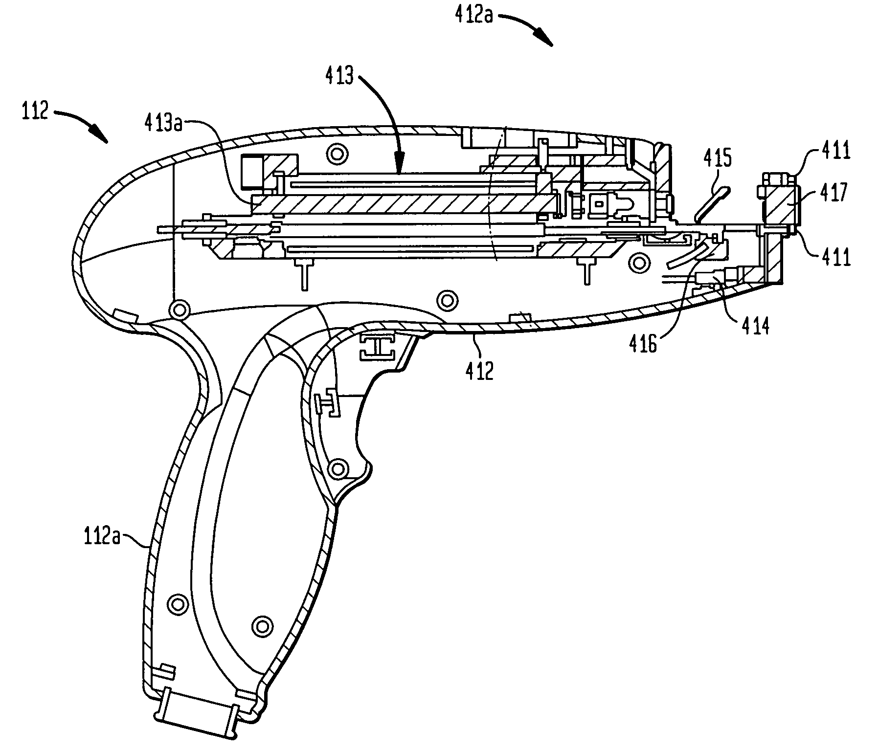
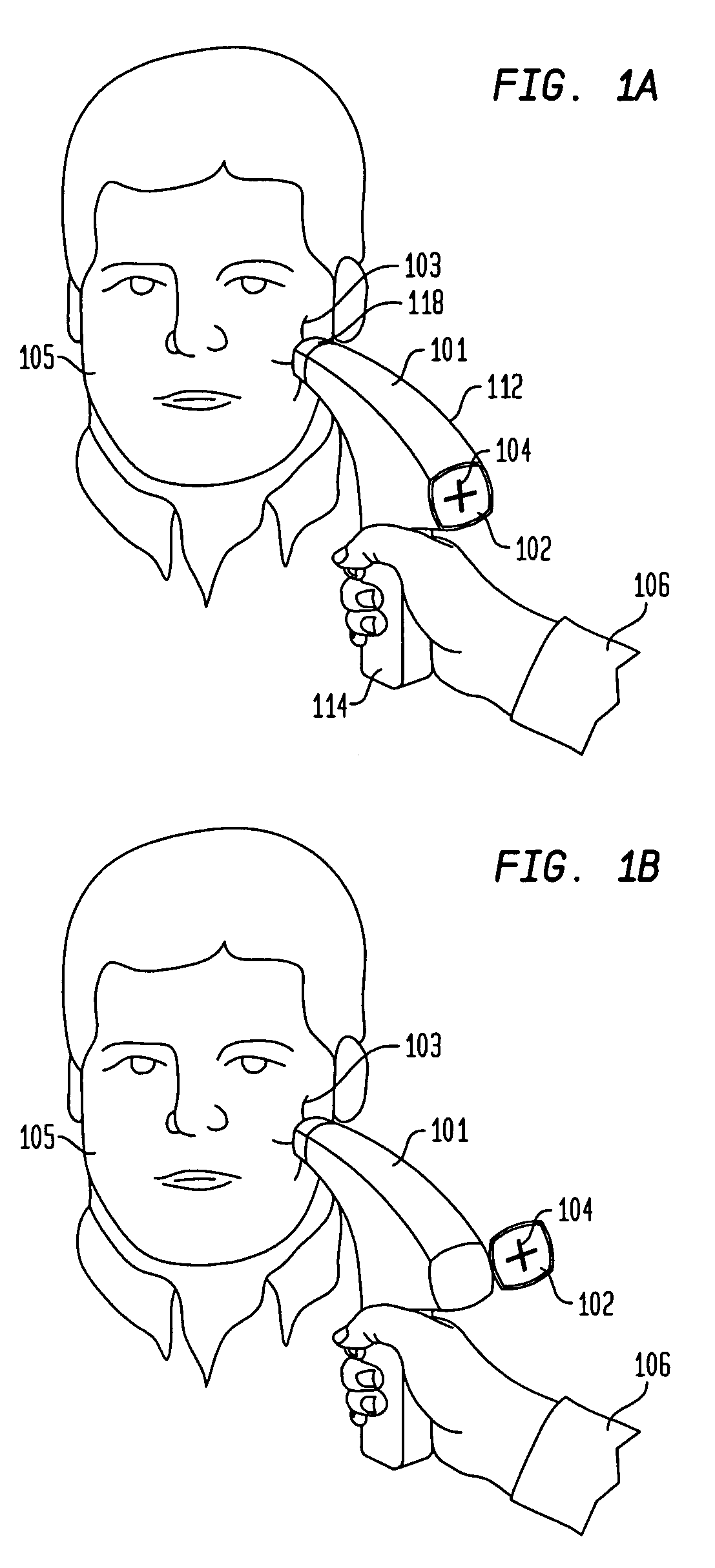
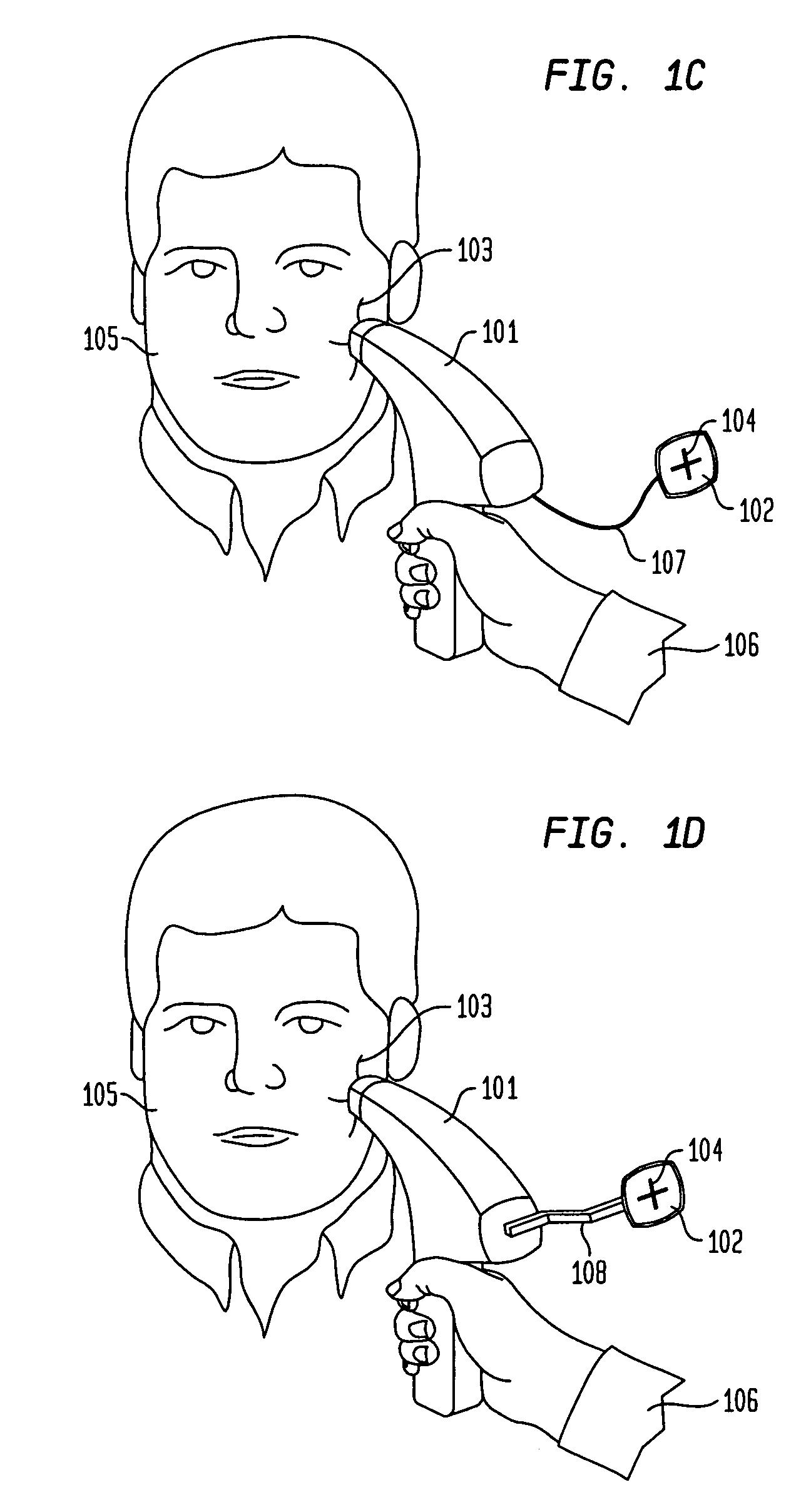
[0083]FIG. 1A schematically depicts a handpiece device 112 according to one embodiment of the invention having a housing 101 that includes a handle 114 that allows a user 106, e.g., a medical professional or a home user, to hold and aim the device at a selected target treatment area 103. The housing, which defines an enclosure in which various components of the device are incorporate...
PUM
 Abstract
Description
Claims
Application Information