

Crystal structure of cytochrome P450
a technology of cytochrome p450 and crystal structure, which is applied in the field of human cytochrome p450 protein 3a4, can solve the problems of chancy and difficult process of crystallising a protein, no clear expectation of success, and the major obstacle in the process of crystallizing protein molecules from solution
- Summary
- Abstract
- Description
- Claims
- Application Information
AI Technical Summary
Benefits of technology
Problems solved by technology
Method used
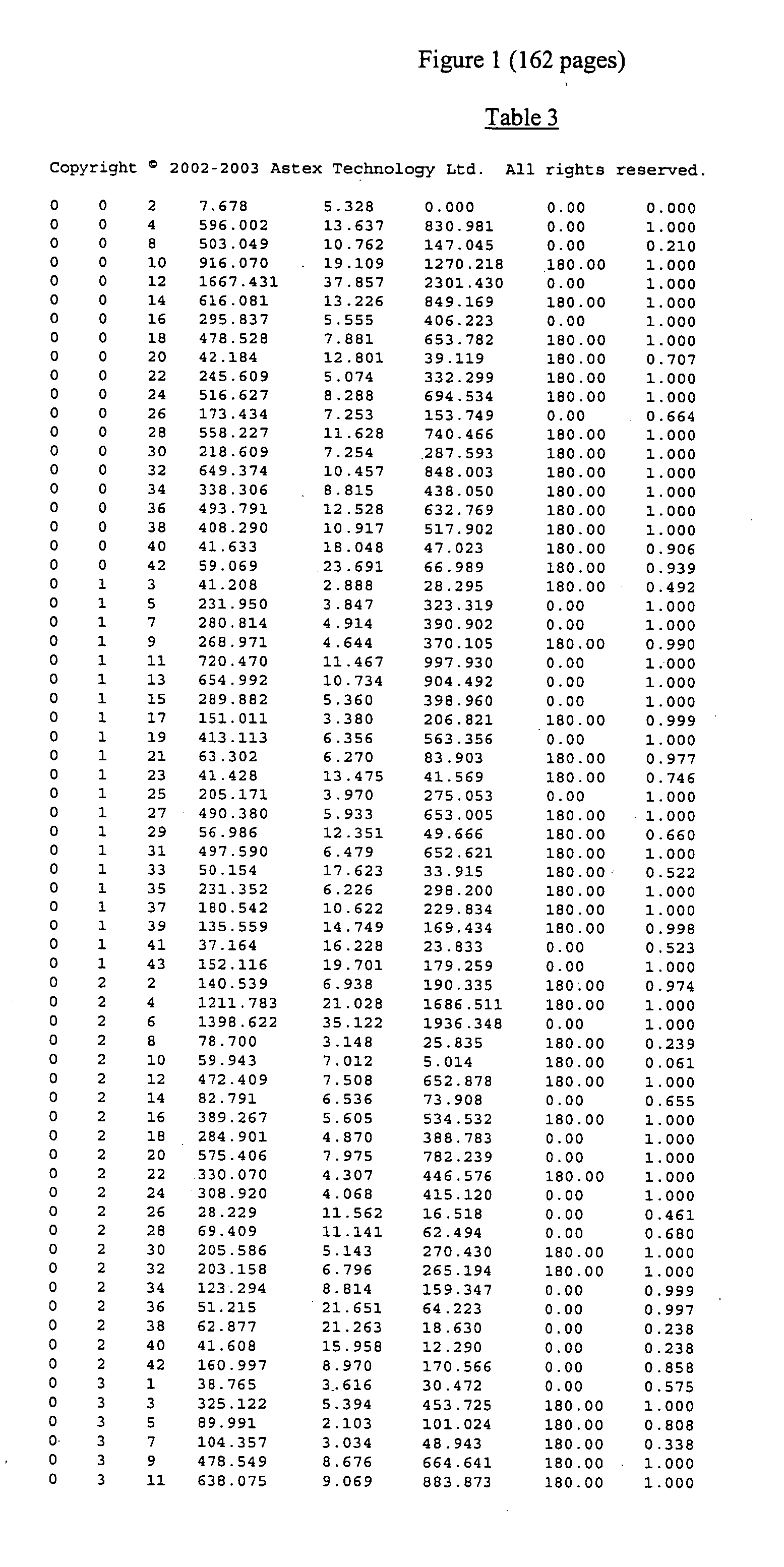
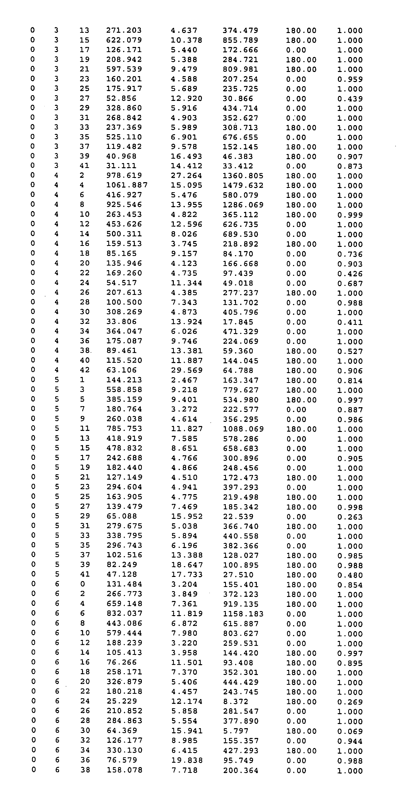
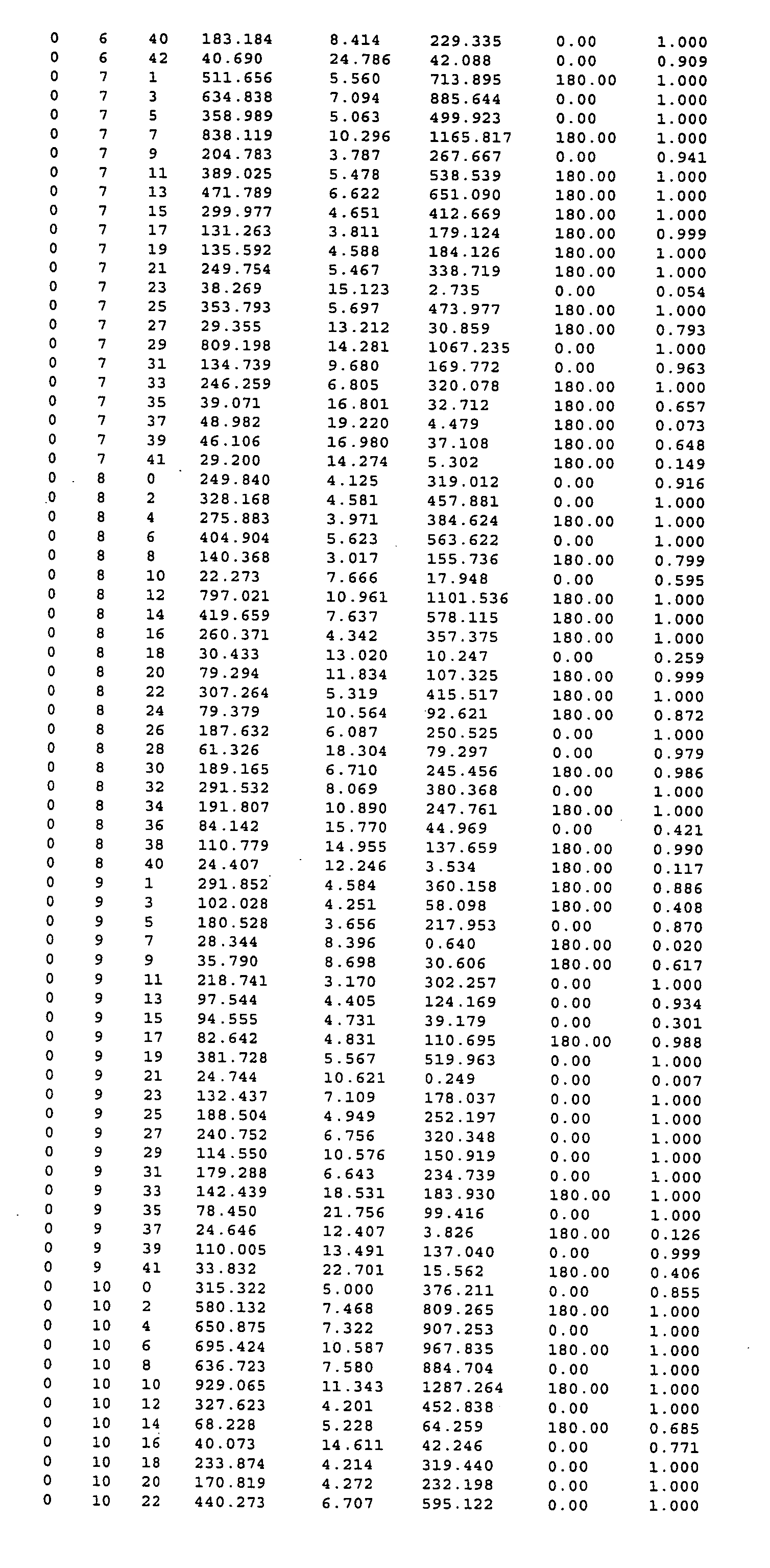
Image

Examples
example
Cloning of 3A4
[0335] 3A4 corresponding to M18907 (GI—181373) was cloned from human liver library (Origene Technologies, Inc.).
[0336] PCR carried out as recommended by the manufacturer:
Liver library 2.0 μl10X PCR buffer (−Mg2+) 2.5 μl10 mM dNTPs 0.5 μl10 mM MgSO4 2.5 μlWater11.0 μlPrimer 1 (@10 pmol / μl) 3.0 μlPrimer 2 (@10 pmol / μl) 3.0 μl
[0337] Primer 1 is complementary to the 5′ end of the full length 3A4 cDNA. Primer 2 is complementary to the 3′ end of the cDNA and adds a four histidine tag onto the C-terminus of the 3A4 protein.
[0338] Heat to 94° C., add 0.5 μl (I Unit) Vent polymerase.
[0339] 35 cycles as follows:
94° C.30 seconds65° C.60 seconds72° C.60 seconds
[0340] 1 cycle of 72° C. for 5 minutes.
[0341] Following the addition of 1 μl (2.5 Units) Taq polymerase and incubation at 72° C. for 10 minutes, 1 μl of product was used in a TOPO cloning reaction (vector pCR4TOPO, Invitrogen). The cloning reaction was used to transform E. coli XL1-blue and positive clones identifi...
PUM
| Property | Measurement | Unit |
|---|---|---|
| concentration | aaaaa | aaaaa |
| molecular weight | aaaaa | aaaaa |
| molecular weight | aaaaa | aaaaa |
Abstract
Description
Claims
Application Information