Eosinophil Major Basic Protein as a natural heparanase-inhibiting protein, compositions, methods and uses thereof
a technology of eosinophils and basic proteins, applied in the field of natural heparanase inhibitors, can solve the problem that none of the prior art heparanase inhibitors are natural proteins
- Summary
- Abstract
- Description
- Claims
- Application Information
AI Technical Summary
Benefits of technology
Problems solved by technology
Method used
Image
Examples
example 1
[0179] Evaluation of Heparanase Expression and Localization in Eosinophils
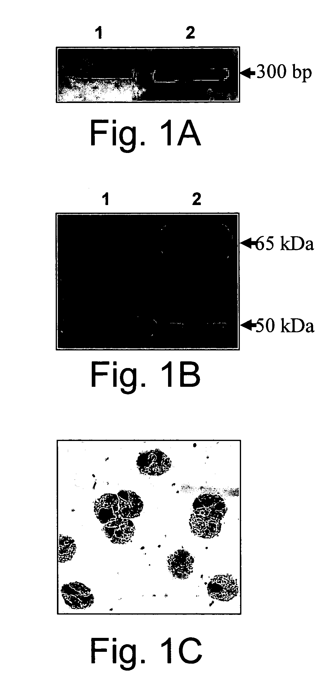
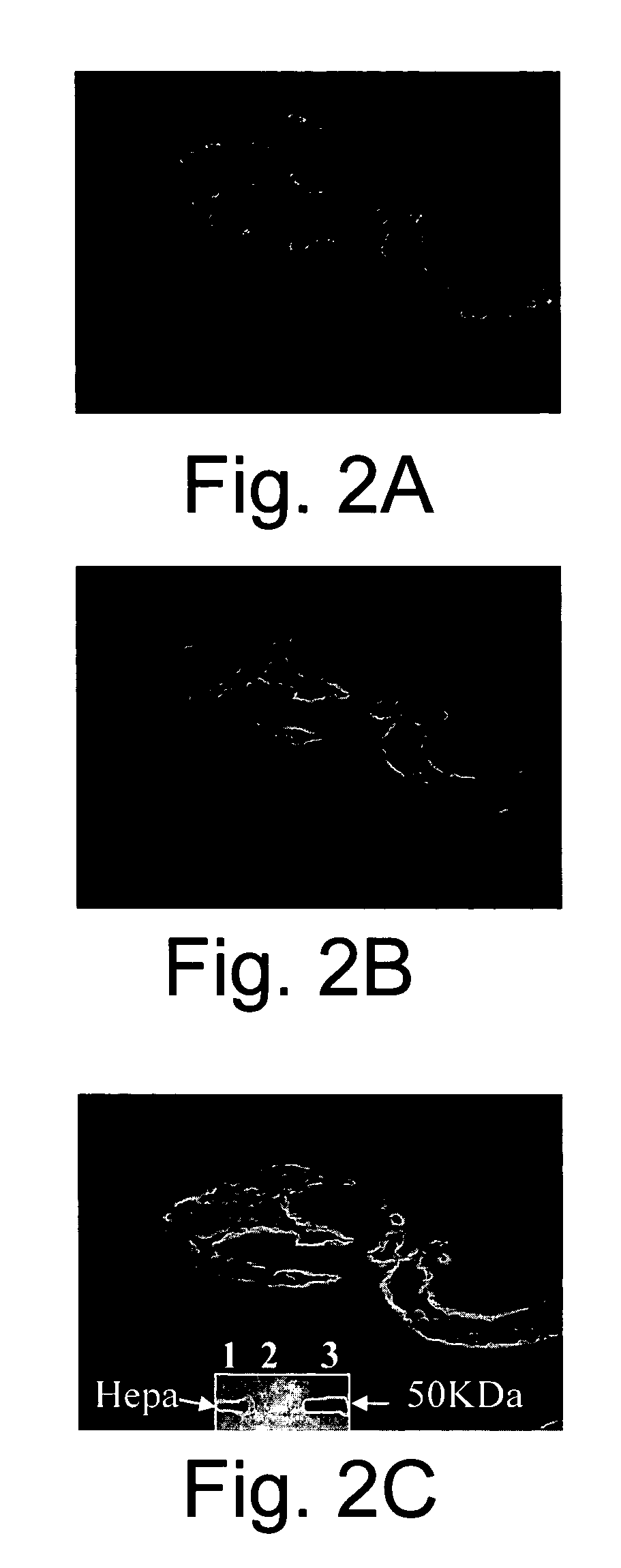
[0180] The increased significant role ascribed recently for heparanase in inflammation, angiogenesis and cancer progression, have led the inventors to investigate the possible involvement of heparanase in inflammation and allergy processes mediated by eosinophils. As a first step, the expression of heparanase mRNA in freshly isolated human peripheral blood eosinophils was demonstrated by RT-PCR using heparanase specific primers (FIG. 1A). Both the processed (50 kDa) and, to a lesser extent, the unprocessed (65 kDa) forms of heparanase were detected by Western blot analysis of lysed freshly isolated eosinophils (FIG. 1B, lane 1). On the basis of this experiment, the inventors estimated that 1×10⁶ eosinophils contain 2-4 ng of heparanase similar to other cells of the immune system [Vlodavsky (1992) ibid.; Matzner (1985) ibid.]. By staining the eosinophils with anti-human heparanase monoclonal antibodies it was ...
example 2
[0183] Evaluation of Eosinophil-Associated Heparanase Enymatic Activity
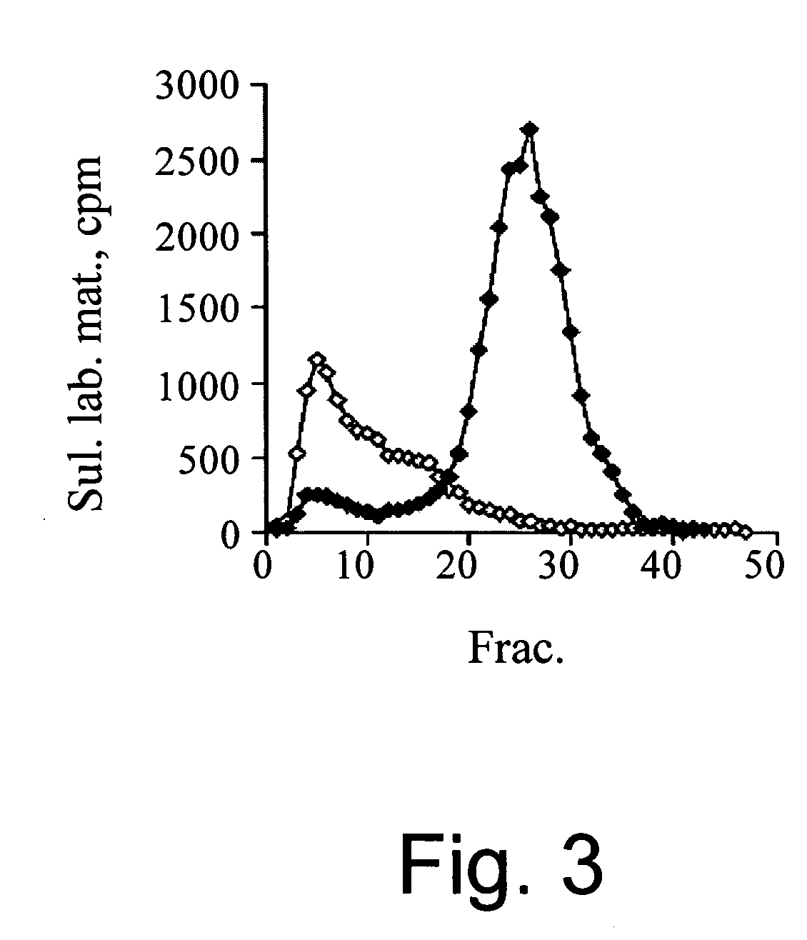
[0184] Next, the inventors evaluated whether eosinophils display heparanase enzymatic activity. For this purpose, lysates of freshly isolated eosinophils were incubated with sulfate labeled ECM. These samples failed to release sulfate labeled HS degradation fragments (FIG. 3), indicating a lack of heparanase enzymatic activity. In contrast, lysed neutrophils exhibited a high heparanase activity, releasing 60-70% of the total ECM incorporated radioactivity in the form of HS degradation fragments (fractions 20-35) (FIG. 3). The biochemical nature of these cleavage fragments was characterized in previous studies [Matzner (1985) ibid.]. Subsequently, the inventors tried to induce heparanase activity by activating the eosinophils for 15 min. or 18 h, with either PAF, PMA, recombinant human skin α-tryptase, IL-2 or sonicated HMC-1 cells. As observed with resting cells, none of these treatments induced heparanase activ...
example 3
[0185] Inhibition of Heparanase Activity by Eosinophils and MBP
[0186] The inventors next hypothesized that eosinophils might inhibit heparanase activity. Therefore, the ability of eosinophil lysates to inhibit heparanase-mediated degradation of HS in intact ECM was investigated. For this purpose, active 50 kDa recombinant heparanase (10 ng / ml) was incubated with sulfate labeled ECM in the absence or presence of either lysed eosinophils, or, as a control, lysed human foreskin fibroblasts. As shown in FIG. 5A, release of low-molecular weight labeled HS degradation fragments was specifically abolished by eosinophil lysates, but not by fibroblasts. In a subsequent experiment, the eosinophil lysates were incubated with the labeled ECM in the presence of increasing concentrations of recombinant heparanase. Complete inhibition of activity was obtained even at an heparanase concentration of 1 μg / ml, representing an estimated excess of MBP over heparanase of about 2.5 folds (not shown).
[01...
PUM
| Property | Measurement | Unit |
|---|---|---|
| concentration | aaaaa | aaaaa |
| diameter | aaaaa | aaaaa |
| pH | aaaaa | aaaaa |
Abstract
Description
Claims
Application Information
 - R&D
- Intellectual Property
- Life Sciences
- Materials
- Tech Scout
- Unparalleled Data Quality
- Higher Quality Content
- 60% Fewer Hallucinations
Browse by: Latest US Patents, China's latest patents, Technical Efficacy Thesaurus, Application Domain, Technology Topic, Popular Technical Reports.
© 2025 PatSnap. All rights reserved.Legal|Privacy policy|Modern Slavery Act Transparency Statement|Sitemap|About US| Contact US: help@patsnap.com


