Detection of dna methylation

a methylation and detection technology, applied in the field of genetic fingerprinting, can solve the problems of difficult to demonstrate unequivocally, lack of efficient procedures for large-scale assessment of dna methylation at cpg sites around the genome, etc., and achieve the effect of better profiling a victim

Inactive Publication Date: 2005-10-13
THE UNIV OF QUEENSLAND
View PDF4 Cites 9 Cited by
  • Summary
  • Abstract
  • Description
  • Claims
  • Application Information

AI Technical Summary

Benefits of technology

[0015] The terms “methylation signature”, “methylation profile” and “methylome” are used interchangeably throughout the specification and all indicate the methylation status of a cell's or group of cells' genome. They can also be used to determine the expression or absence of expression of particular genes. The method of the present invention is useful in determining methylation polymorphisms which can then be linked to particular traits and / or phenotypic characteristics or stages of aging or development and can also be useful in forensics to assist in better profiling a victim or perpetrator.

Problems solved by technology

The challenge now is to understand how the genome is expressed in a certain fashion and in particular cell types to produce the traits that ultimately determine the nature and quality of life.
A role for DNA methylation in human disease has also been widely suggested but difficult to demonstrate unequivocally.
One of the problems limiting this important field of research has been the lack of efficient procedures for large scale assessment of DNA methylation at CpG sites around the genome.

Method used


Image

  • Detection of dna methylation
  • Detection of dna methylation
  • Detection of dna methylation

Examples


example 1

AMP Protocol

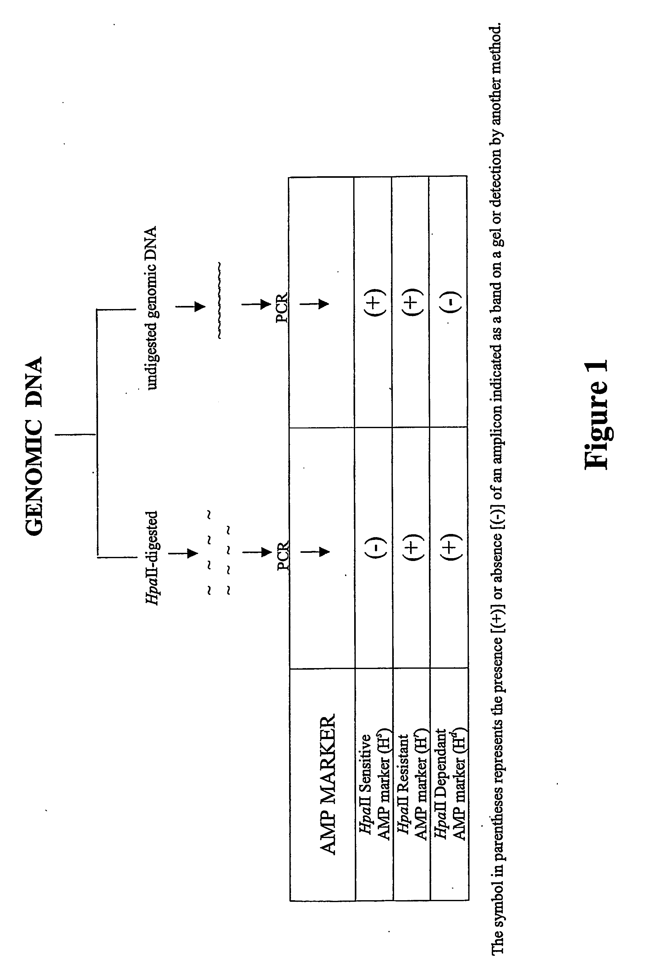
[0129] The following protocol may need to be adapted for use in particular plant or animal cells. However, in general, the procedure involves isolating genomic DNA, subjecting the DNA to HpaII digestion and subjecting the digest to an amplification reaction using a primer which spans all or part of the HpaII recognition sequence, i.e.

 ↓CCGG,   ↑

such that if HpaII has digested the nucleotide sequence, primer extension cannot occur.

[0130] A diagram showing the AMP protocol is shown in FIG. 1.

[0131] Three classes of product are expected:

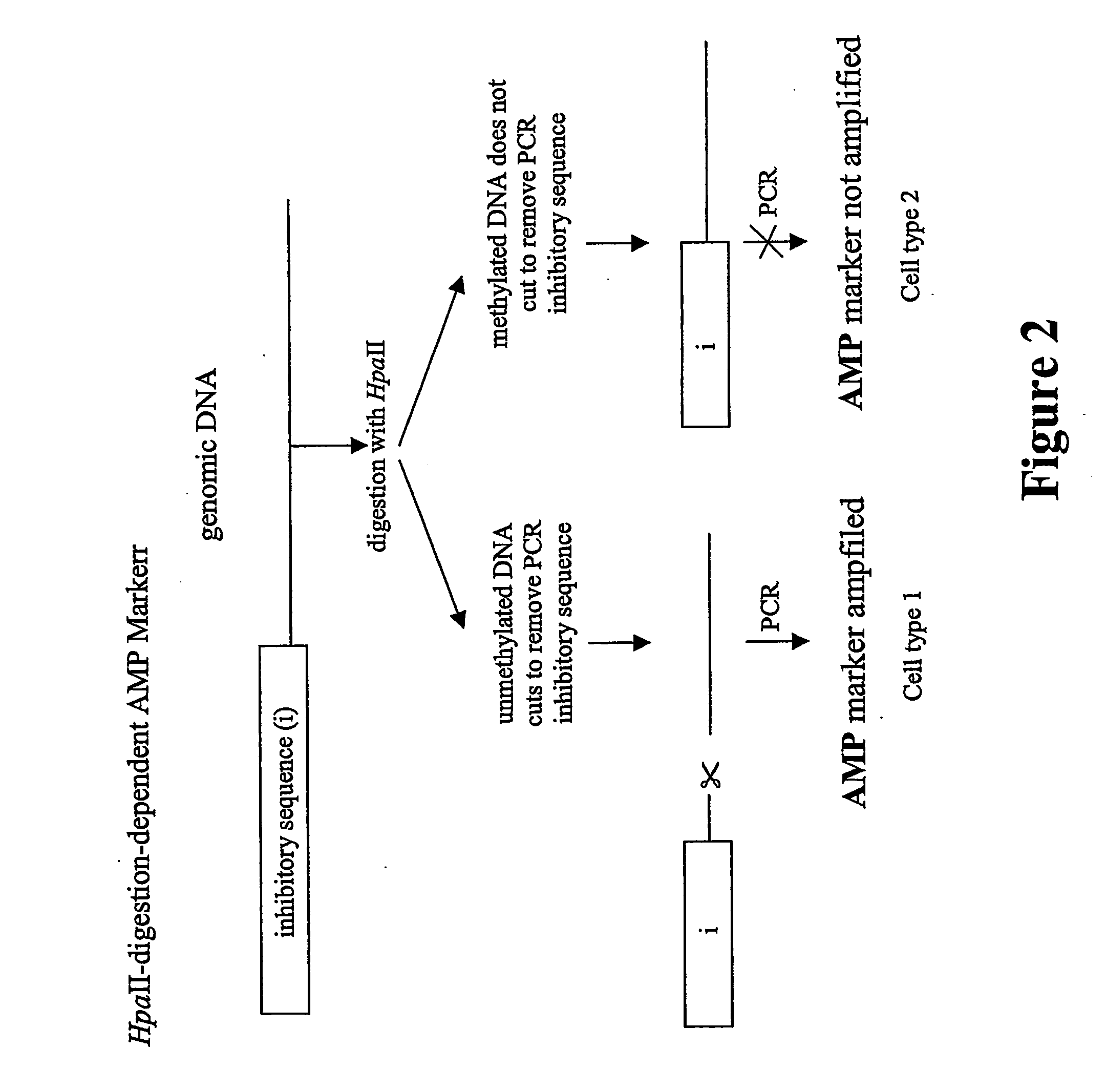
Class IHpaII digestion-resistant (Hr) markers, indicative ofmethylationClass IIHpaII digestion-sensitive (Hs) markers, indicative of non-methylation.Class IIIHpaII digestion-dependent (Hd) markers. Amplicon ismethylated or partly methylated and is linked to anunmethylated HpaII site followed by an inhibitor sequence.Cleavage of the unmethylated site removes inhibition andallows amplification.

[0132] The existence of an Hd (Class III) ...

example 2

Detection of DNA Polymorphisms in Human Body Fluid

[0169] Using the AMP technology, abundant and distinct tissue-specific DNA methylation polymorphisms were readily detected between human blood and sperm (FIG. 4). Most of the tissue specific DNA methylation polymorphisms were Class III digestion dependent markers and surprisingly, 30% of these were polymorphic in methylation status between these tissues. Between five blood samples and three sperm samples (from eight separate individuals), 80% of variation in AMP profiles generated from HapII-digested template could be explained by the tissue type, and the remaining 20% of variation could not be distinguished from nucleotide (rather than DNA methylation) polymorphism.

[0170] In another example, blood from three sets of young, healthy, identical twins were also assessed for DNA methylation polymorphisms over 8,000 CpG sites in the genome, representing an average of about 340 CpG sites per human chromosome. There was 100% concordance o...

example 3

Identification of Class III Markers in Human Genome

[0171]FIG. 5 is a diagrammatic representation showing map location of four human digestion-dependent Class III AMP markers and the flanking HpaII sites. Each of the digestion-dependent markers that was cloned and sequenced was 100% homologous to sequenced regions of the human genome. The positions of the Class III amplicons (HdM 1-4) [SEQ ID NOS:33-36] are indicated by thick shaded boxes on the chromosomal segment. The locations of HpaII sites are represented by vertical lines and the distance to, and orientation of nearby genes are indicated. Southern analysis has shown that Class III markers represent the junction between methylated and unmethylated DNA in the genome. Tissue-specific Class III AMP markers often map to the CpG islands and often represent tissue-specific modification of methylation in or around CpG islands.


PUM

PropertyMeasurementUnit
emission wavelengthaaaaaaaaaa

Abstract

The present invention relates generally to a method of genetic fingerprinting and more particularly to a method of establishing a methylation signature for a particular eukaryotic cell or group of cells such as in the form of tissue cells or organ-specific cells. The method of the present invention exploits the sensitivity of certain restriction enzymes to methylation. Exposure of genomic or transgene DNA to these enzymes followed by amplification results in products which establish the methylation signature or profile (i.e. the methylome profile) of a cell's or group of cells' genome when compared to the amplified genome in undigested form. The method of the present invention is useful inter alia in phenotyping a cell based on its methylation signature and provides a useful tool in functional genomics and for the design of therapeutic and trait-modifying protocols, particularly for animals and plants. The present invention can also be used to identify and map junctions between methylated and unmethylated DNA. The method of the present invention is also useful for identifying DNA methylation polymorphisms which can be used inter alia in diagnosis and forensics and for identifying particular genes, the function or absence of function of which are associated with a disease condition or trait. The method is also useful for monitoring the aging process of particular cells or an animal (including a human) or plant comprising such cells as well as monitoring the pluripotent or multipotent state of stem cells and development of stem cells through to maturation.

Description

FIELD OF THE INVENTION [0001] The present invention relates generally to a method of genetic fingerprinting and more particularly to a method of establishing a methylation signature for a particular eukaryotic cell or group of cells such as in the form of tissue cells or organ-specific cells. The method of the present invention exploits the sensitivity of certain restriction enzymes to methylation. Exposure of genomic or transgene DNA to these enzymes followed by amplification results in products which establish the methylation signature or profile (i.e. the methylome profile) of a cell's or group of cells' genome when compared to the amplified genome in undigested form. The method of the present invention is useful inter alia in phenotyping a cell based on its methylation signature and provides a useful tool in functional genomics and for the design of therapeutic and trait-modifying protocols, particularly for animals and plants. The present invention can also be used to identify ...

Claims


Application Information

Patent Timeline
Patent Type & Authority Applications(United States)
IPC IPC(8): C12Q1/68C12Q1/683
CPCC12Q1/683C12Q2521/331C12Q2531/113
Inventor CARROLL, BERNARDHARRISON, DIONAUNG, HNIN
Owner THE UNIV OF QUEENSLAND
Why Eureka
Try Eureka