Detection of acute myocardial infarction biomarkers

a biomarker and myocardial infarction technology, applied in the field of early detection of acute myocardial infarction, can solve the problems of permanent heart muscle damage, unnecessarily admitting 2.5 million patients with low-risk chest pain to the hospital as a precaution, and spending 5 billion or more of unnecessary medical costs

Inactive Publication Date: 2005-11-10
NEOMECS
View PDF16 Cites 118 Cited by
  • Summary
  • Abstract
  • Description
  • Claims
  • Application Information

AI Technical Summary

Benefits of technology

[0016] One embodiment relates to a device for detecting biomarker analytes indicative of acute myocardial infarction or drug resistance in a fluid sample. The device includes an optical material body having a surface-textured area. A plasma polymerized layer is associated with the surface-textured area on the optical material body. An analyte-specific chemistry is coupled to the plasma polymerization layer, the analyte specific chemistry being specific for a biomarker analyte indicative of acute myocardial infarction or drug resistance. The analyte-specific chemistry has at least one optical property sensitive to binding of the biomarker analyte.
[0017] A further embodiment relates to a device for detecting biomarker analytes indicative of acute myocardial infarction or drug resistance in a fluid sample. The device includes an optical material body having a first surface-textured area and a second surface-textured area. A plasma polymerized layer is associated with the first surface-textured area and a plasma polymerized layer is associated with the second surface-textured area on the optical material body. An analyte-specific chemistry is coupled to the plasma polymerization layer associated with the first surface-textured area and an analyte-specific chemistry is coupled to the plasma polymerized layer associated with the second surface-textured area, the analyte specific chemistry for associating with a biomarker analyte indicative of acute myocardial infarction or drug resistance. The analyte-specific chemistry has at least one optical property sensitive to binding of the biomarker analyte thereto.
[0018] Another embodiment relates to a method for detecting acute myocardial infarction biomarkers or drug resistance in a patient. An optical material body having a textured surface and having elongated projections, a plasma polymerization-modified surface, and at least one analyte-specific chemistry is obtained. A fluid sample is placed on the optical material body. The fluid sample is separated into a plurality of fluid components on the optical material body, and at least one of the components contains analytes. The separated fluid component containing analytes is placed adjacent the elongated projections of the textured surface on the optical material body such that the separated component is received within the elongated projections. The separated fluid component within the elongated projections is optically sensed to detect analyte biomarkers for myocardial infarction or drug resistance.
[0019] Another embodiment includes a method for making an optical element for detecting impending myocardial infraction or drug resistance. The optical material body is etched with atomic oxygen to obtain a textured surface. A plasma polymerized layer is adhered to the textured surface by plasma polymerization. An analyte-specific chemistry is adhered to the plasma polymerized layer, the analyte-specific chemistry being specific for a biomarker analyte indicative of either acute myocardial infarction or drug resistance. The analyte-specific chemistry has at least one optical property sensitive to binding of the biomarker analyte thereto.

Problems solved by technology

In addition, U.S. government statistics indicate that $5 billion or more of unnecessary medical costs are spent each year on the assessment of non-cardiac (i.e., false positive) cases in hospital emergency departments.
Approximately 2.5 million low-risk chest pain patients are unnecessarily admitted to the hospital as a precaution and at a significant cost to the U.S. healthcare system.
In addition, only fifty percent of the patients experiencing ACS receive appropriate therapy in a timely fashion, and this delayed therapy can result in permanent heart muscle damage and greater total cost for patient care.
Acute plaque rupture is highly recognized as the primary cause of acute thrombus formation and complete occlusion of the vessel may result in irreversible ischemic damage to the cardiac tissue supplied downstream from the obstruction.
Often the patient does not exhibit complete occlusion of the vessel but has a substantial lesion (greater than 90%) and thus is at extremely high risk of complete blockage and eventual acute myocardial infarction.
However, serious cardiac events must be “ruled-out,” such that patients at risk of an eventual AMI are not released.
It is difficult to separate cardiac from non-cardiac events for the millions of people presenting with chest pain.
Currently, no tests are available in a readily usable format that allow for the rapid and specific determination of platelet, pro-coagulation, or pro-inflammatory biomarkers.
Attempts to develop a reproducible test to indicate platelet activation have encountered two significant difficulties.
The second difficulty is that platelets then need to be separated from the withdrawn blood by centrifugation, which also can activate the platelets.
Consequently, testing results may not reflect a patient's true or authentic platelet activation status.

Method used


Image

  • Detection of acute myocardial infarction biomarkers
  • Detection of acute myocardial infarction biomarkers
  • Detection of acute myocardial infarction biomarkers

Examples


Embodiment Construction

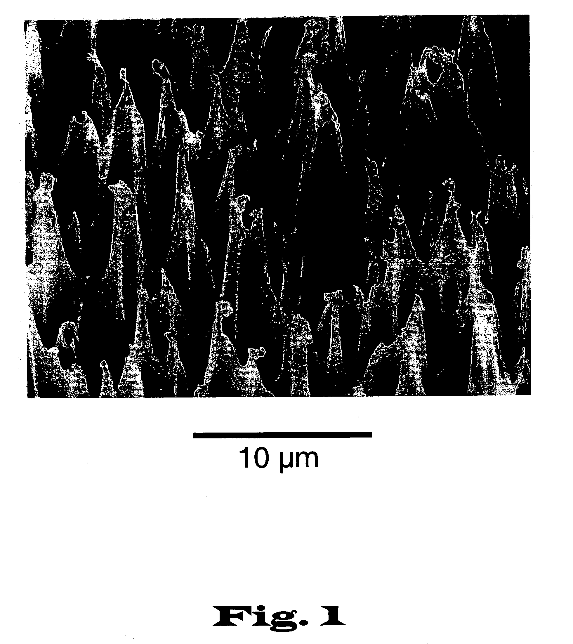
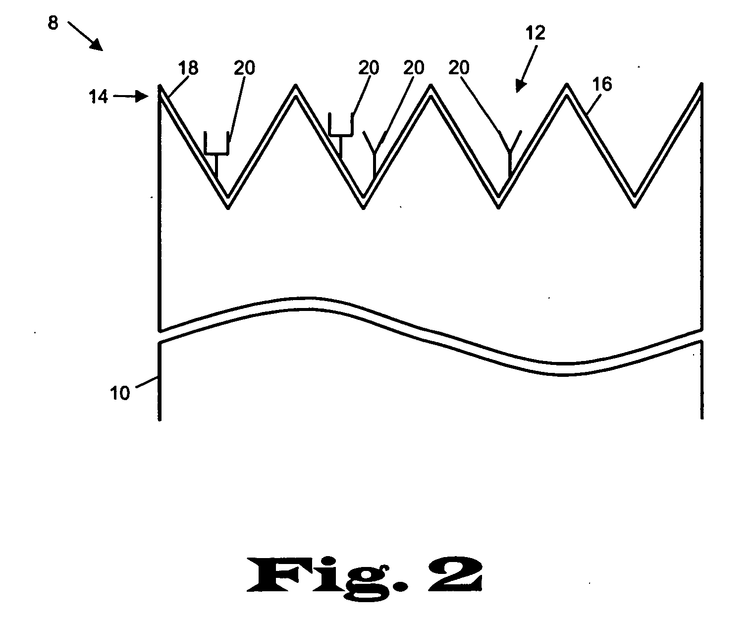
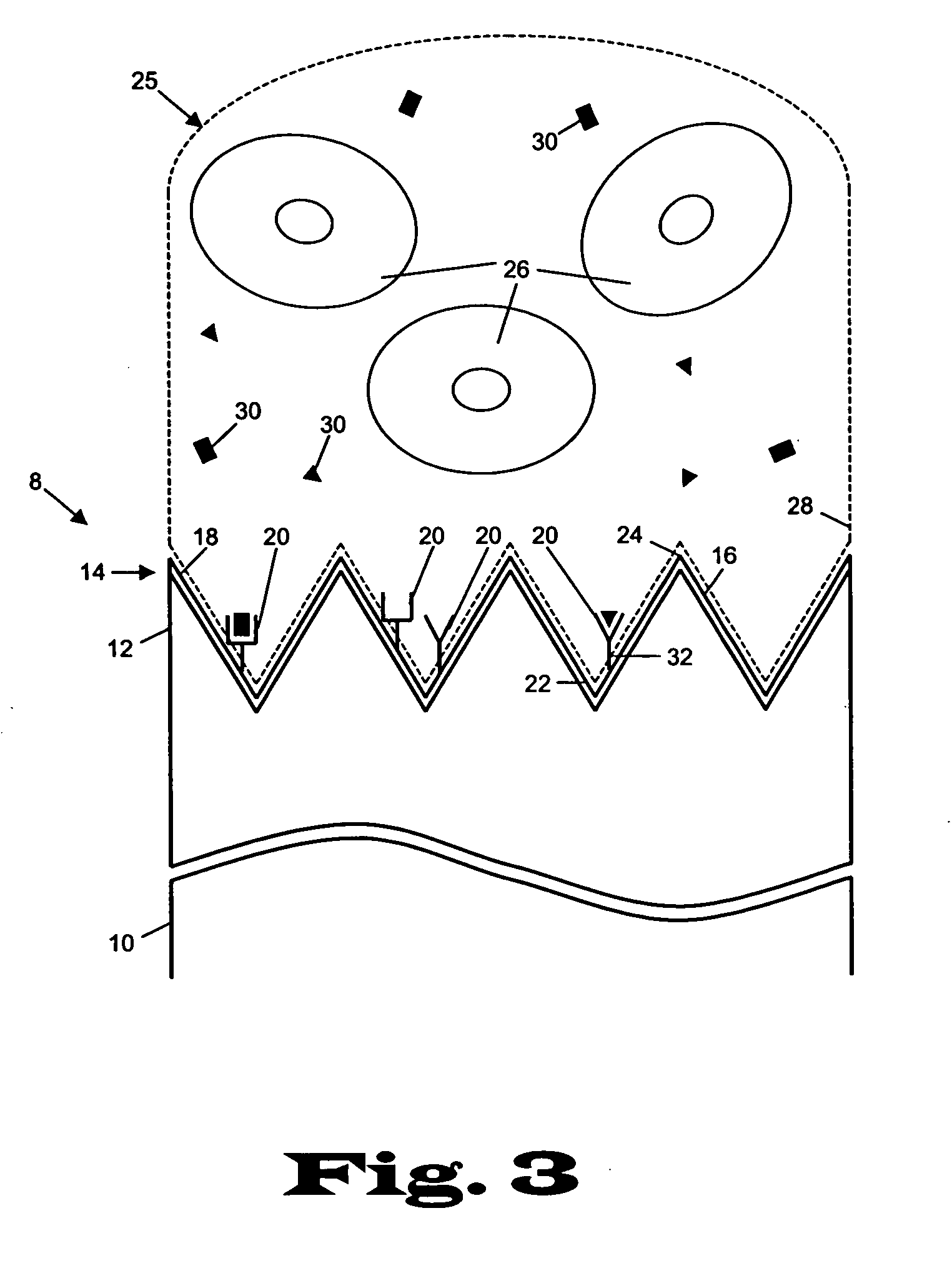
[0023] The present invention relates to devices and methods for the analysis of biological fluid samples, such as blood, for acute myocardial infarction (AMI) precursors or biomarkers, using a biosensor technology. While reference will be made to blood throughout, the fluid sample can include other biological samples, such as urine or saliva. The sensor provides for the spatial separation of the cellular elements of the blood, and provides a rapid analysis of the separated blood plasma component using reagents attached to the sensor, which are specific to the biomarker being measured. Therapeutic cardiovascular drug monitoring can also be performed with the assays. These assays can measure specific platelet and coagulation proteins that participate early in the evolution of a thrombus (blood clot) and later in a potential acute myocardial infarction (AMI). By assaying a blood sample for these AMI precursors / biomarkers, a determination can be made much earlier whether the presenting ...


PUM

PropertyMeasurementUnit
diameteraaaaaaaaaa
diameteraaaaaaaaaa
diameteraaaaaaaaaa

Abstract

The present invention relates to medical devices and methods for early detection of acute myocardial infarction in a patient. In particular, the invention relates to a device and method for detecting the presence of biomarker analytes in a specimen which are indicative of a potential acute myocardial infarction in the patient.

Description

CROSS-REFERENCE TO RELATED APPLICATIONS [0001] This application claims the benefit of U.S. Provisional Patent Application Ser. No. 60 / 516,655 filed Oct. 31, 2003 (Shebuski et al., “Detection of Acute Myocardial Infarction Precursors”), Ser. No. 60 / 516,656 filed Oct. 31, 2003 (Nomura, “Method and Apparatus for Body Fluid Analysis Using Surface-Textured Optical Materials”), and U.S. Provisional Patent Application Ser. No. 60 / 516,654 filed Oct. 31, 2003 (Nomura, “Plasma Polymerization of Atomically Modified Surfaces”), which hereby are incorporated herein by reference thereto in their entirety.FIELD OF THE INVENTION [0002] The present invention relates to medical devices and diagnostic methods for the early detection of acute myocardial infarction in a patient. In particular, the invention relates to a device and method for timely and sensitive detection of specific analytes in a fluid sample, such as a blood specimen which may be indicative of a potential or impending acute myocardial...

Claims


Application Information

Patent Timeline
Patent Type & Authority Applications(United States)
IPC IPC(8): A61KC12M1/34C12M3/00C23CG01NG01N21/00G01N33/53G01N33/543G01N33/573G01N33/68G02B6/00
CPCB05D1/62C08J7/047C08J7/18C08J2333/00C08J2433/00H05H1/24G01N33/54366G01N33/6893G01N2800/32G01N2800/324G01N33/54353C08J7/0427
Inventor SHEBUSKI, RONALD JOHNKYDD, ARTHUR RAYMONDNOMURA, HIROSHI
Owner NEOMECS
Why Eureka
Try Eureka