
Method and apparatus for imaging of tissue using multi-wavelength ultrasonic tagging of light

a multi-wavelength, ultrasonic technology, applied in the field of non-invasive optical imaging, can solve the problems of low or reduced sensitivity and specificity of imaging techniques, poor spatial resolution and anatomical registration of optical imaging,

Inactive Publication Date: 2005-11-17
GENERAL ELECTRIC CO
View PDF17 Cites 47 Cited by
  • Summary
  • Abstract
  • Description
  • Claims
  • Application Information

AI Technical Summary

Benefits of technology

[0005] In accordance with certain implementation of the present technique, an exemplary imaging apparatus is disclosed. The imaging apparatus includes a source of electromagnetic radiation that is configured to emit electromagnetic radiation at two or more wavelengths through an imaging volume. The imaging apparatus also includes an ultrasound transducer that is ...

Problems solved by technology

Due to light absorption and scattering by the imaged tissue, optical imaging typically has relatively poor spatial resolution and anatomical registration.
For example, when optical imaging is employed for cancer detection, the imaging technique suffers from low or reduced sensitivity and specificity.

Method used


Image

  • Method and apparatus for imaging of tissue using multi-wavelength ultrasonic tagging of light
  • Method and apparatus for imaging of tissue using multi-wavelength ultrasonic tagging of light
  • Method and apparatus for imaging of tissue using multi-wavelength ultrasonic tagging of light

Examples


Embodiment Construction

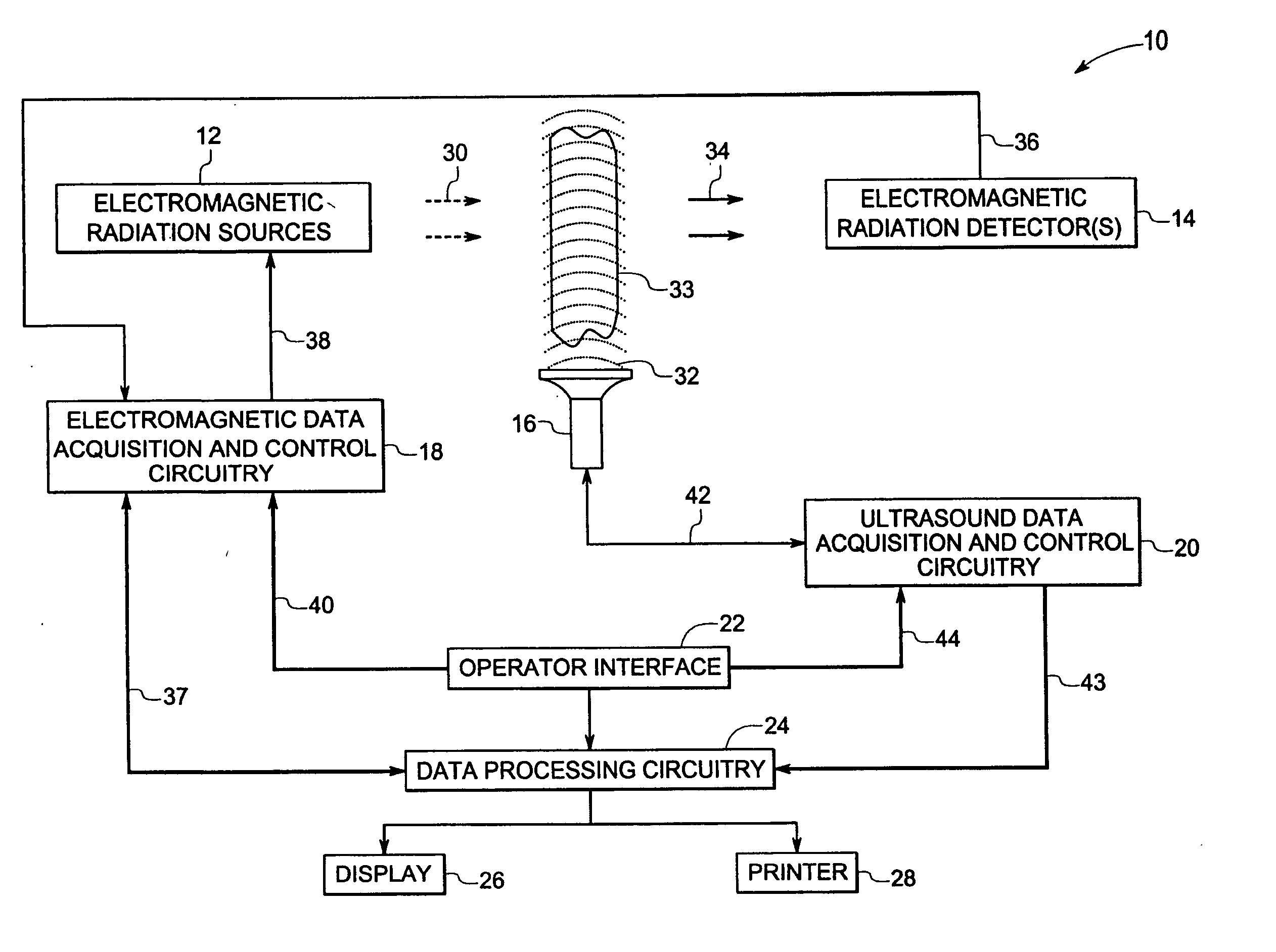
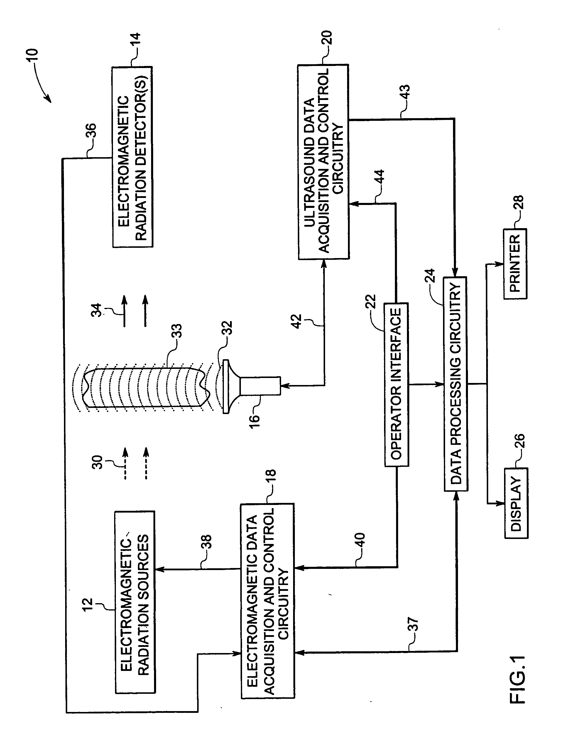
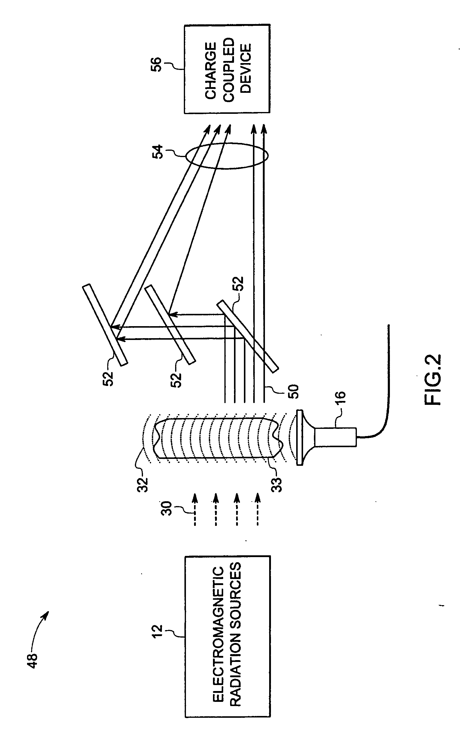
[0010] Turning now to the drawings and referring first to FIG. 1, an exemplary imaging system 10 is illustrated. The depicted imaging system 10 includes a source 12 of electromagnetic radiation, a detector 14 of electromagnetic radiation, an ultrasound transducer 16, electromagnetic data acquisition and control circuitry 18, ultrasound data acquisition circuitry 20, operator interface 22, and data processing circuitry 24. The depicted exemplary imaging system 10 also includes a display 26 and a printer 28. The source 12 and detector 14 define an imaging volume which may accommodate part of a patient undergoing imaging, such as tissues 33 or organs being examined for signs of a disease or disorder.

[0011] The source 12 is configured to emit electromagnetic radiation 30 at two or more wavelengths. In one embodiment, the wavelengths are in a range from about 600 nanometers to about 900 nanometers, which is typically classified as near-infrared (NIR) radiation. As will be appreciated by...


PUM


Abstract

An imaging apparatus is disclosed. The imaging apparatus includes a source of electromagnetic radiation that is configured to emit electromagnetic radiation at two or more wavelengths through an imaging volume. The imaging apparatus also includes an ultrasound transducer that is configured to direct ultrasound waves through the imaging volume such that electromagnetic radiation passing through the imaging volume is modulated at the frequency of the ultrasound waves. The imaging apparatus further includes one or more electromagnetic radiation detectors that are configured to detect the modulated electromagnetic radiation from the imaging volume.

Description

CROSS REFERENCE TO RELATED APPLICATIONS [0001] The present application claims priority to the provisional patent application No. 60 / 570645, filed on May 12, 2004, and entitled “Method And Apparatus For Functional Imaging Of Biological Tissue Using Multi-Wavelength Ultrasonic Tagging Of Light”.BACKGROUND [0002] The invention relates generally to the field of diagnostic medical imaging and more specifically, to non-invasive optical imaging. [0003] In modern healthcare facilities, non-invasive imaging systems are often used for identifying, diagnosing, and treating physical conditions. Medical imaging typically encompasses the different non-invasive techniques to image and visualize the internal structures and / or functional behavior (such as chemical or metabolic activity) of organs and tissues within a patient. Currently, a number of modalities exist for medical diagnostic and imaging systems, each typically operating on different physical principles to generate different types of ima...

Claims


Application Information

Patent Timeline
IPC IPC(8): A61B5/00A61B8/00A61B8/14
CPCA61B5/0073A61B2562/0238A61B8/14A61B5/0097
Inventor FOMITCHOV, PAVEL ALEXEYEVICH
Owner GENERAL ELECTRIC CO
Features
  • Generate Ideas
  • Intellectual Property
  • Life Sciences
  • Materials
  • Tech Scout
  • Unparalleled Data Quality
  • Higher Quality Content
  • 60% Fewer Hallucinations
Learn More