
Methods and apparatuses for conducting assays

a technology of assays and apparatuses, applied in the field of methods for conducting assays of samples, can solve the problems of difficult comparison of results with well established reference ranges that have been expressed, limited acceptance of assays carried out in whole blood, and difficulty in carrying out precise and accurate measurements

Active Publication Date: 2006-01-26
MESO SCALE TECH LLC
View PDF10 Cites 84 Cited by
  • Summary
  • Abstract
  • Description
  • Claims
  • Application Information

AI Technical Summary

Benefits of technology

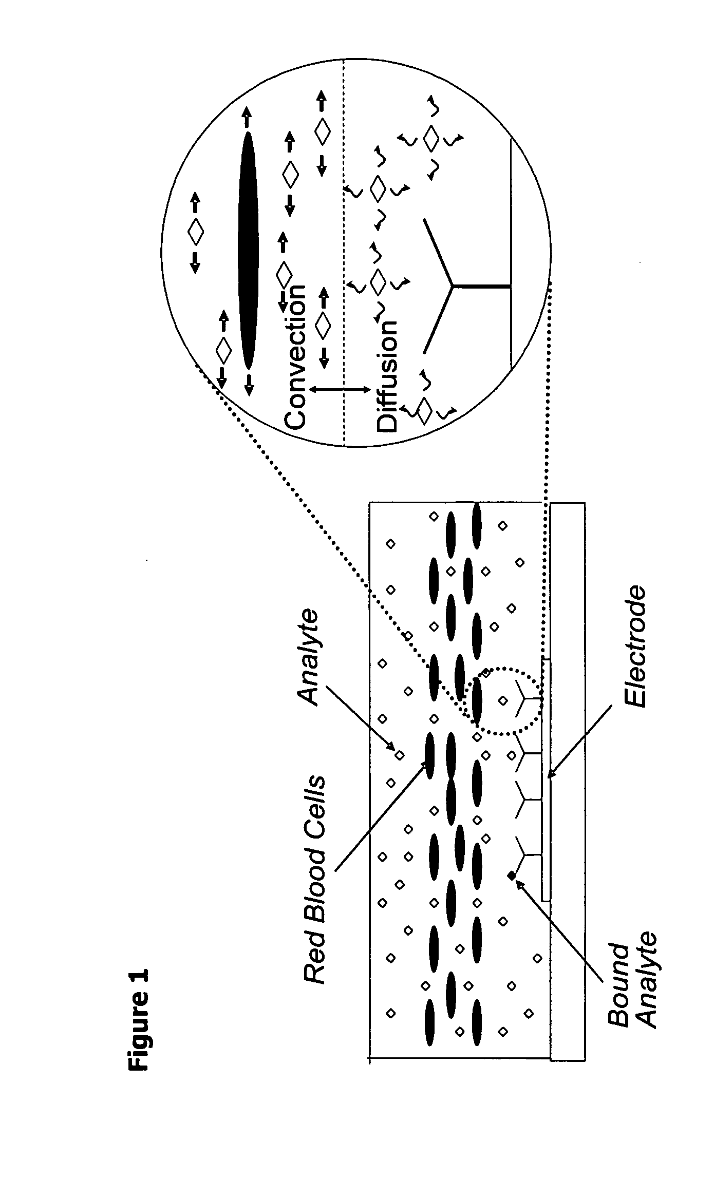
The invention relates to methods for measuring analytes in samples containing particulate matter, such as whole blood samples. The methods allow for the measurement of the concentration of analytes in the liquid fraction of the sample without removing the particulate matter or correcting for the volume occupied by the particulate matter. The measured concentration closely approximates the actual concentration of the analyte in the sample. The invention also includes methods for measuring the concentration of analytes in the fluid fraction of a sample, which is independent of the hematocrit of the sample. The act of contacting at least a portion of a sample with a surface is also a method of the invention.

Problems solved by technology

Assays carried out in whole blood have had limited acceptance, in part because conventional assays give different signals for a whole blood sample relative to a plasma or serum fraction derived from the same sample.
The difference in signals makes it difficult to compare results to well established reference ranges that have been expressed in terms of the concentration of the analyte in serum or plasma.
In addition, the presence of a large volume fraction of cells in a whole blood sample may also interfere with many assay technologies and make it difficult to carry out precise and accurate measurements.
Because of the wide range for hematocrit values that may be observed in patient samples (the normal range is from 41-50% for adult males and 35-46% for adult females, but hematocrit levels of less than 20% can be observed in severe cases of anemia), this approach could lead to large errors in the reported values.
This approach adds complexity to the assay apparatus and may be subject to increased imprecision because the error is a function of the variability in both the assay signal and hematocrit determination.
While these approaches avoid the need for a hematocrit correction, they add significant cost and complexity to the instrumentation.
In addition, filtration has the disadvantages of possible loss of analyte to the filter material and limitations in the sample volume that can be easily processed.

Method used


Image

  • Methods and apparatuses for conducting assays
  • Methods and apparatuses for conducting assays
  • Methods and apparatuses for conducting assays

Examples


example 1

Hematocrit-Independent One-Step Sandwich Immunoassay for Plasma Levels of Cardiac Markers in Whole Blood Samples

[0108] Six test samples having the same plasma levels of cardiac markers but different hematocrit levels were prepared as described in Materials and Methods. Table 1 provides, for each of the samples, the hematocrit, the concentration of each of the analytes in the blood sample (blood concentration) and the concentration of each of the analytes in the plasma fraction (plasma concentration).

TABLE 1HematocritBlood Concentration (ng / mL)Plasma Concentration (ng / mL)Sample(%)MyoTnICKMBTnTMyoTnICKMBTnT102002.3183.12012.3183.12191631.9152.52012.3183.13281451.7132.32012.3183.14371271.4112.22012.3183.15471071.29.51.62012.3183.1656881.07.91.42012.3183.1

[0109] A solution containing detection antibodies against myoglobin, TnI, CKMB and TnT (3.3 μl) was mixed with 160 μl of each test sample. The final antibody concentrations (in weight per total blood volume) were 2 μg / ml with the ex...

example 2

Hematocrit-Independent One-Step Sandwich Immunoassay for Plasma Levels of Cardiac Markers in Whole Blood Samples

[0111] Example 1 compared the signals obtained for whole blood samples that varied in hematocrit but contained equal levels of analyte in their plasma fractions. In this example, whole blood samples are analyzed that have equal concentrations of analyte with respect to the total volume of blood. The concentrations of analyte in the plasma should, therefore, vary as a function of the sample hematocrit.

[0112] The stock blood sample was prepared by combining 2282 μl of blood (hematocrit level of 42.5%) with 51 μl of a mixture of detection antibodies for myoglobin, cTnT, cTnI, and CKMB at concentrations 193, 97, 163, and 98 μg / ml, respectively and 33 μl of the BSA and bovine IgG blocking agent solution. Whole blood samples at four hematocrit levels were prepared from stock blood sample as described in the Materials and Methods except that the blood stock contained no added a...

example 5

Hematocrit-Independent Two-Step Sandwich Immunoassay for Plasma Levels of Cardiac Markers in Whole Blood Samples

[0118] Whole blood samples with equal plasma levels of cardiac markers but varying hematocrits were prepared as in Example 4. The detection antibody solution had 5 ug / mL of labeled anti-TnT and 3 ug / mL of anti-CKMB. The detection antibody solution also contained anti-TnI and anti-myoglobin detection antibodies; the number of labels per antibody for these two antibodies was, however, too low to get a good signal in the TnI and CKMB assays and these data are not presented.

[0119] The assays were conducted in two-step format. FIG. 8 shows the normalized signal. As observed in the analogous one step assay (Example 1), the signals were independent of hematocrit.


PUM

PropertyMeasurementUnit
Reynold's numberaaaaaaaaaa
Reynold's numbersaaaaaaaaaa

Abstract

Disclosed are methods for conducting assays of samples, such as whole blood, that may contain cells or other particulate matter. Also disclosed are systems, devices, equipment, kits and reagents for use in such methods. One advantage of certain disclosed methods and systems is the ability to rapidly measure the concentration of an analyte of interest in blood plasma from a whole blood sample without blood separation and hematocrit correction.

Description

RELATED APPLICATIONS [0001] This application claims the benefit of priority to: U.S. Provisional Patent Application Ser. No. 60 / 576,710, filed Jun. 3, 2004, titled METHODS AND APPARATUSES FOR CONDUCTING ASSAYS, by M. Billadeau, et al., which is incorporated herein by reference.FIELD OF THE INVENTION [0002] The present invention is directed to methods for conducting assays of samples, such as whole blood, that may contain cells or other particulate matter. The invention also relates to systems, devices, equipment, kits and reagents for use in such methods. BACKGROUND OF THE INVENTION [0003] Traditionally, clinical measurements (especially, solid phase binding assays) for analytes in blood have been carried out in serum or plasma samples derived from the blood. Partly for this reason, clinicians present the levels of most blood markers in terms of the concentration of the marker in the liquid fraction of the blood sample (e.g., the concentration in plasma or serum fractions derived fr...

Claims


Application Information

Patent Timeline
Patent Type & Authority Applications(United States)
IPC IPC(8): G01N33/567G01N33/52G01N33/543
CPCG01N33/523G01N33/543G01N33/54366Y10T436/106664Y10T436/25375Y10T436/117497Y10T436/25125
Inventor BILLADEAU, MARKDEBAD, JEFFGLEZER, ELILELAND, JONATHANWIJAYAWARDHANA, CHARLES
Owner MESO SCALE TECH LLC
Learn More