Methods for electrosurgical tissue treatment between spaced apart electrodes

a tissue treatment and electrode technology, applied in the field of electrosurgical tissue treatment between spaced apart electrodes, can solve problems such as tissue necrosis to a depth, achieve the effects of reducing power delivery and ablation rate, limiting tissue necrosis depth, and increasing power delivery with electrosurgical devices

Inactive Publication Date: 2006-02-23
ARTHROCARE
View PDF99 Cites 14 Cited by
  • Summary
  • Abstract
  • Description
  • Claims
  • Application Information

AI Technical Summary

Benefits of technology

[0013] The use of such electrode arrays in electrosurgical procedures is particularly advantageous as it has been found to limit the depth of tissue necrosis without substantially reducing power delivery and ablation rates. Heretofore, increased power delivery with electrosurgical devices has generally been achieved by increasing monolithic electrode area. The resulting large electrode surfaces, however, cause tissue necrosis to a depth which varies proportionally with the width and area of the electrode surface. The present invention provides a more controlled necrosis depth by utilizing a plurality of isolated electrode terminals, where the terminals are preferably laterally spaced-apart by a distance from one-tenth to one terminal diameter, with spacing between larger electrode terminals generally being at the lower end of the range. Such spacing provides adequate power delivery and ablation rates without excessive tissue necrosis, which is usually limited to a depth less than one electrode terminal diameter.

Problems solved by technology

The resulting large electrode surfaces, however, cause tissue necrosis to a depth which varies proportionally with the width and area of the electrode surface.

Method used


Image

  • Methods for electrosurgical tissue treatment between spaced apart electrodes
  • Methods for electrosurgical tissue treatment between spaced apart electrodes
  • Methods for electrosurgical tissue treatment between spaced apart electrodes

Examples


Embodiment Construction

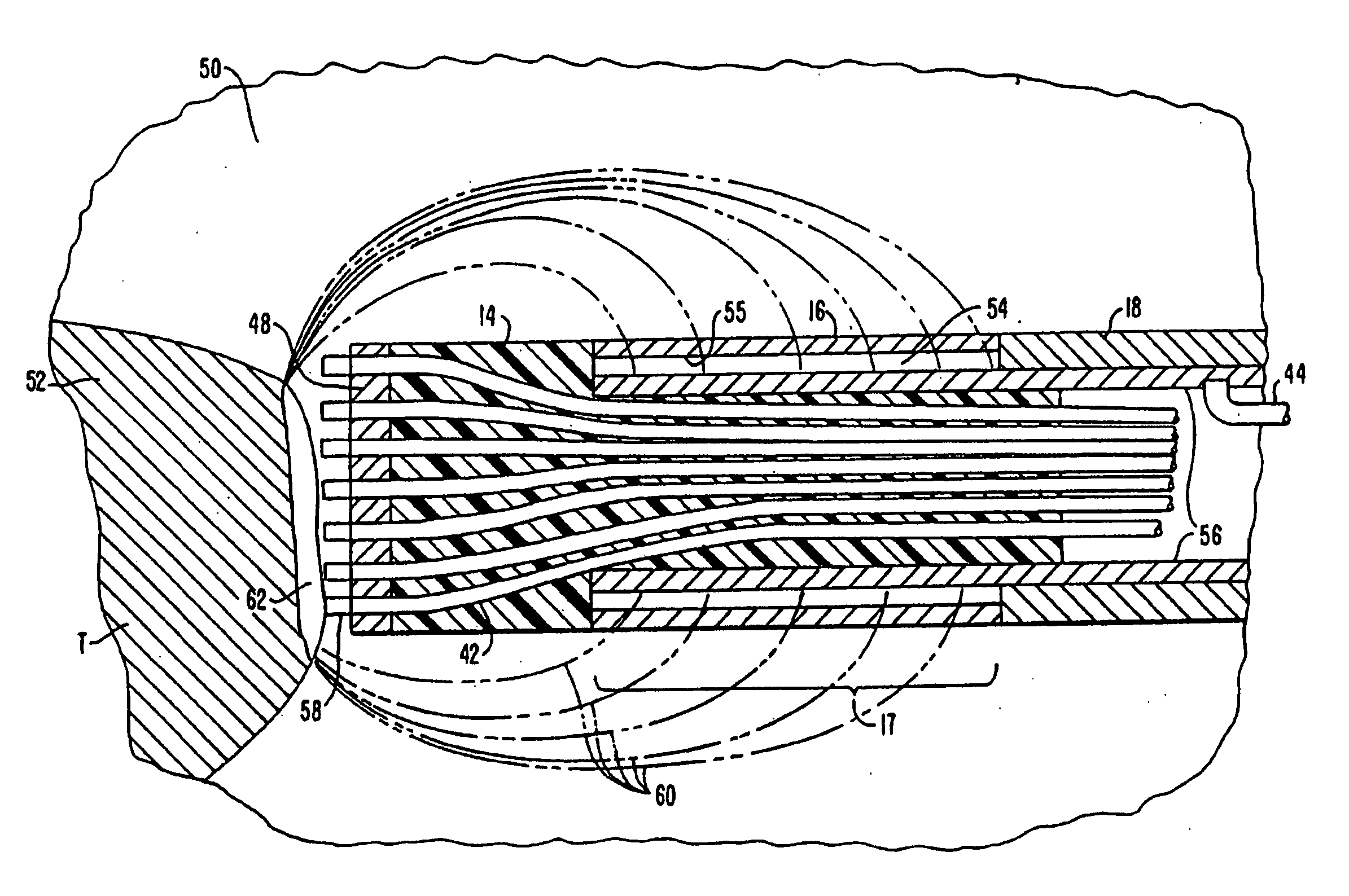
[0039] The present invention provides a method and apparatus for selectively heating a target location within a patient's body, such as solid tissue or the like, particularly including articular cartilage, fibrocartilage, meniscal tissue, and the like. In addition to articular cartilage and fibrocartilage, tissues which may be treated by the method and apparatus of the present invention include tumors, abnormal tissues, and the like. For convenience, the remaining disclosure will be directed specifically at the cutting, shaping or ablation of fibrocartilage and articular cartilage during arthroscopic or endoscopic procedures but it will be appreciated that the apparatus and methods can be applied equally well to procedures involving other tissues of the body, as well as to other procedures including open surgery, laparoscopic surgery, thoracoscopic surgery, and other endoscopic surgical procedures.

[0040] The target tissue will be, by way of example but not limited to, articular car...


PUM


Abstract

As electrosurgical prove comprises a shaft having an electrode array (12) at its distal end and a connector at its proximal end. The array (12) includes a plurality of isolated electrode terminals, and an electrosurgical power supply (28) is provided with a multiplicity of independently limited or controlled current sources and a connector. The electrosurgical probe and the power supply may be connected through their respective connectors so that the independent current sources are connected to individual electrode terminals. By applying very high frequency electrical energy to the electrode array, target tissue may be cut or ablated while heat dissipation through low impedance paths, such as blood and normal saline, will be minimized.

Description

BACKGROUND OF THE INVENTION [0001] 1. Field of the Invention [0002] The present invention relates generally to the field of electrosurgery and, more particularly, to surgical devices and methods which employ very high frequency electrodes comprising an array of individual, isolated electrode terminals. [0003] The field of electrosurgery includes a number of loosely related surgical techniques which have in common the application of electrical energy to modify the structure or integrity of patient tissue. Electrosurgical procedures usually operate through the application of very high frequency currents to cut or ablate tissue structures, where the operation can be monopolar or bipolar. Monopolar techniques rely on external grounding of the patient, where the surgical device defines only a single electrode pole. Bipolar devices comprise both electrodes for the application of current between their surfaces. [0004] Electrosurgical procedures and techniques are particularly advantageous ...

Claims


Application Information

Patent Timeline
Patent Type & Authority Applications(United States)
IPC IPC(8): A61B18/14A61B17/00A61B17/22A61B18/00A61B18/12A61B19/00A61F2/02A61M25/01A61N1/05A61N1/40
CPCA61B18/12A61N1/403A61B18/1402A61B18/148A61B18/1482A61B18/1485A61B18/149A61B18/1492A61B2017/00026A61B2017/00084A61B2017/00097A61B2017/00101A61B2017/00247A61B2017/00292A61B2017/003A61B2017/22001A61B2017/22038A61B2018/00029A61B2018/0016A61B2018/00178A61B2018/00196A61B2018/00392A61B2018/00577A61B2018/00583A61B2018/00601A61B2018/00642A61B2018/0066A61B2018/00678A61B2018/00726A61B2018/00761A61B2018/00791A61B2018/00797A61B2018/00821A61B2018/00827A61B2018/00875A61B2018/00886A61B2018/1213A61B2018/124A61B2018/126A61B2018/1273A61B2018/1407A61B2018/1467A61B2018/1472A61B2018/162A61B2019/5276A61B2218/002A61B2218/007A61F2/2493A61M25/0133A61M25/0147A61M25/0158A61N1/05A61B18/1206A61B2090/378
Inventor EGGERS, PHILIP E.THAPLIYAL, HIRA V.
Owner ARTHROCARE
Why Eureka
Try Eureka