

Method for displaying virtual slide and terminal device for displaying virtual slide
a terminal device and virtual slide technology, applied in the field of virtual slide and terminal device for displaying virtual slide, can solve the problems of difficult re-classification work, disadvantageous restriction of examination work in a specified room,
- Summary
- Abstract
- Description
- Claims
- Application Information
AI Technical Summary
Benefits of technology
Problems solved by technology
Method used
Image

Examples
Embodiment Construction
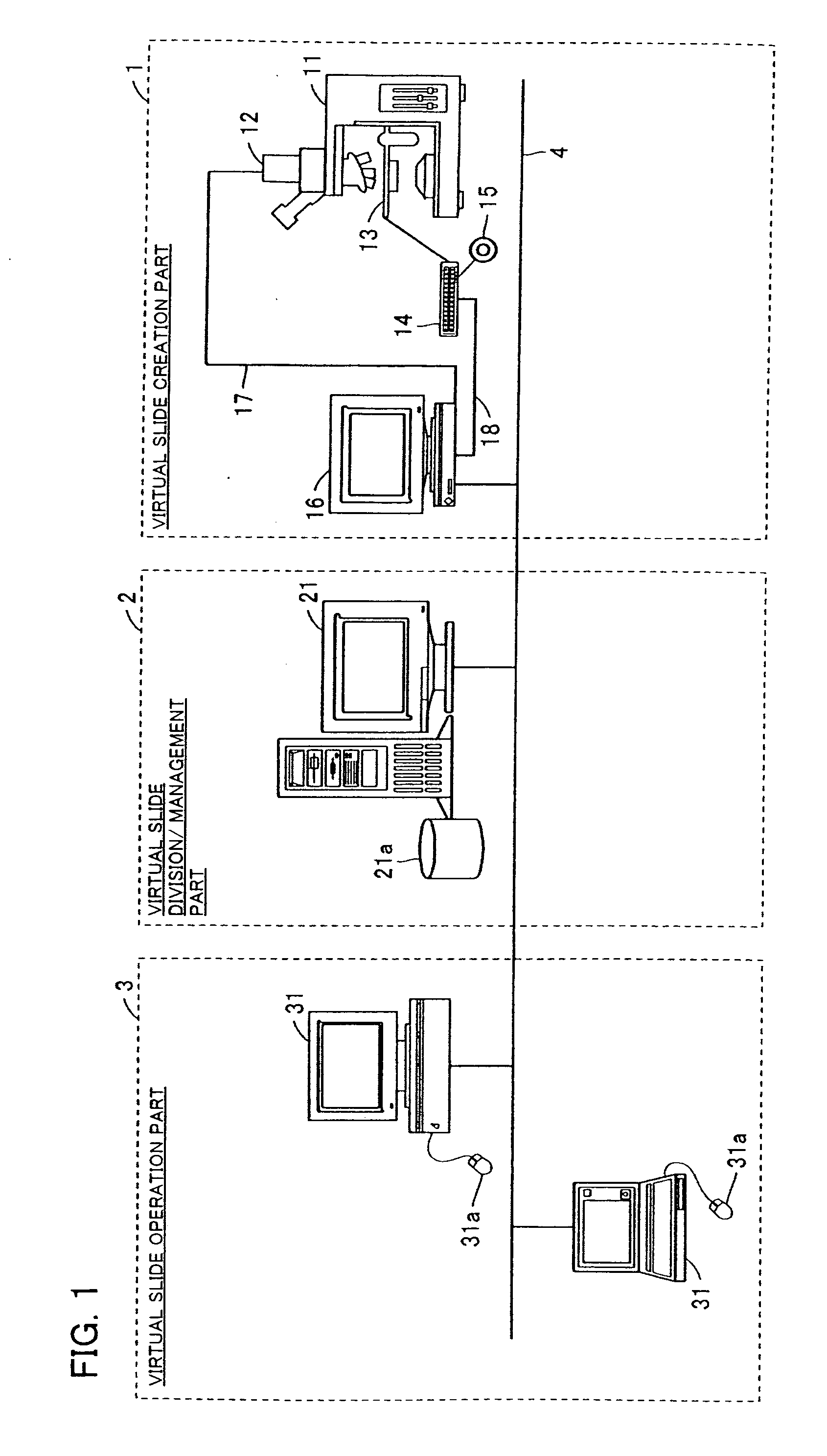
[0032]FIG. 1 shows an overall system construction for practical application of the display method of the virtual microscope slide (blood cell image) according to an embodiment of the invention.
[0033] The system for practically applying the virtual slide display method according to this embodiment is composed of a virtual slide creation part 1, a virtual slide division and management part 2, and a virtual slide operation part 3 as shown in FIG. 1. The virtual slide creation part 1 comprises an optical microscope 11 for confirming the sample slide having magnifications of 20 and 100; a 3CCD camera 12 for incorporating images; an automatic stage 13 for positional control of the optical microscope 11 in X, Y and Z directions; a control unit 14 and a joystick 15 for controlling the automatic stage 13; and an automatic stage control terminal 16 for controlling the automatic stage 13 as well as for focus synthesis and image tiling. The optical microscope 11 used is, for example, a BX-50 s...
PUM
 Abstract
Description
Claims
Application Information