Method and apparatus for imaging of vessel segments

a technology of vessel segments and imaging methods, applied in the field of method and apparatus for imaging of vessel segments, can solve the problems of limiting the ability of patients to quantify the risk, no method other than optical coherence tomography, and no reliable identification of microscopic features that characterize vulnerable plaques. the effect of diagnosis, treatment and prevention

Active Publication Date: 2006-03-16
THE GENERAL HOSPITAL CORP
View PDF99 Cites 143 Cited by
  • Summary
  • Abstract
  • Description
  • Claims
  • Application Information

AI Technical Summary

Benefits of technology

[0015] It is therefore one of the objects of the present invention to provide an apparatus and method that combine quality imaging of internal surfaces of blood vessels and other biological structures and effective imaging of segments of the internal surfaces of the blood vessels. Another object of the present invention is to provide an apparatus and method that provide quality images of internal surfaces of segments of blood vessels in order to offer an improved understanding of the natural history and clinical significance of these lesions which will accelerate progress in diagnosis, treatment and prevention of coronary artery disease.

Problems solved by technology

Although autopsy studies have been valuable in determining features of culprit plaques, the retrospective nature of these studies may limit their ability to quantify the risk of an individual plaque for causing acute coronary thrombosis.
Unfortunately, the microscopic features that characterize vulnerable plaque are not reliably identified by the conventional imaging technologies, such as intravascular ultrasound (“IVUS”), catscan (“CT”), and magnetic resonance imaging (“MRI”).
While experimental intracoronary imaging modalities such integrated backscatter IVUS, elastography, angioscopy, near-infrared spectroscopy, Raman spectroscopy and thermography have been investigated for the detection of vulnerable plaque, it is believed that no method other than optical coherence tomography (“OCT”) has been shown to reliably identify the characteristic features of these lesions.
Strong attenuation of light in blood may present a significant challenge for intravascular optical imaging methods.
However, the combination of the limited flush duration and low image acquisition rate may reduce comprehensive coronary screening.
While this procedure is commonly conducted in Japan, the potential for coronary dissection and myocardial ischemia precludes widespread clinical application of this procedure.
Although these compounds may hold promise for future clinical application, they are not yet approved for human use.
Image quality degrades rapidly in such case, however, manifested by a decrease in transverse resolution as can be seen in FIG. 1A.
As can be clearly seen, the image quality of Image A far exceeds the image quality of Image B. This degradation is unacceptable for most clinical applications.
For technical reasons specific to the current OCT paradigm, an increase in A-line rate may result in an unacceptable penalty in signal to noise ratio, and thus, images of sufficient quality for accurate diagnosis cannot be obtained.

Method used


Image

  • Method and apparatus for imaging of vessel segments
  • Method and apparatus for imaging of vessel segments
  • Method and apparatus for imaging of vessel segments

Examples


Embodiment Construction

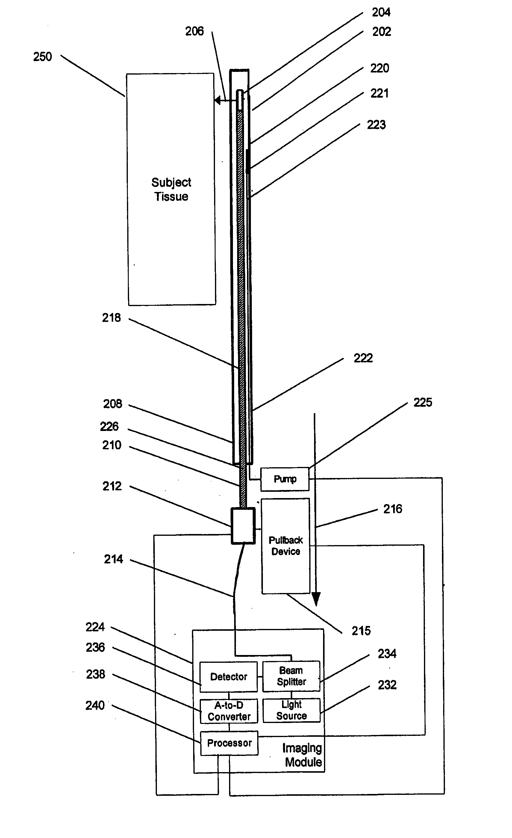
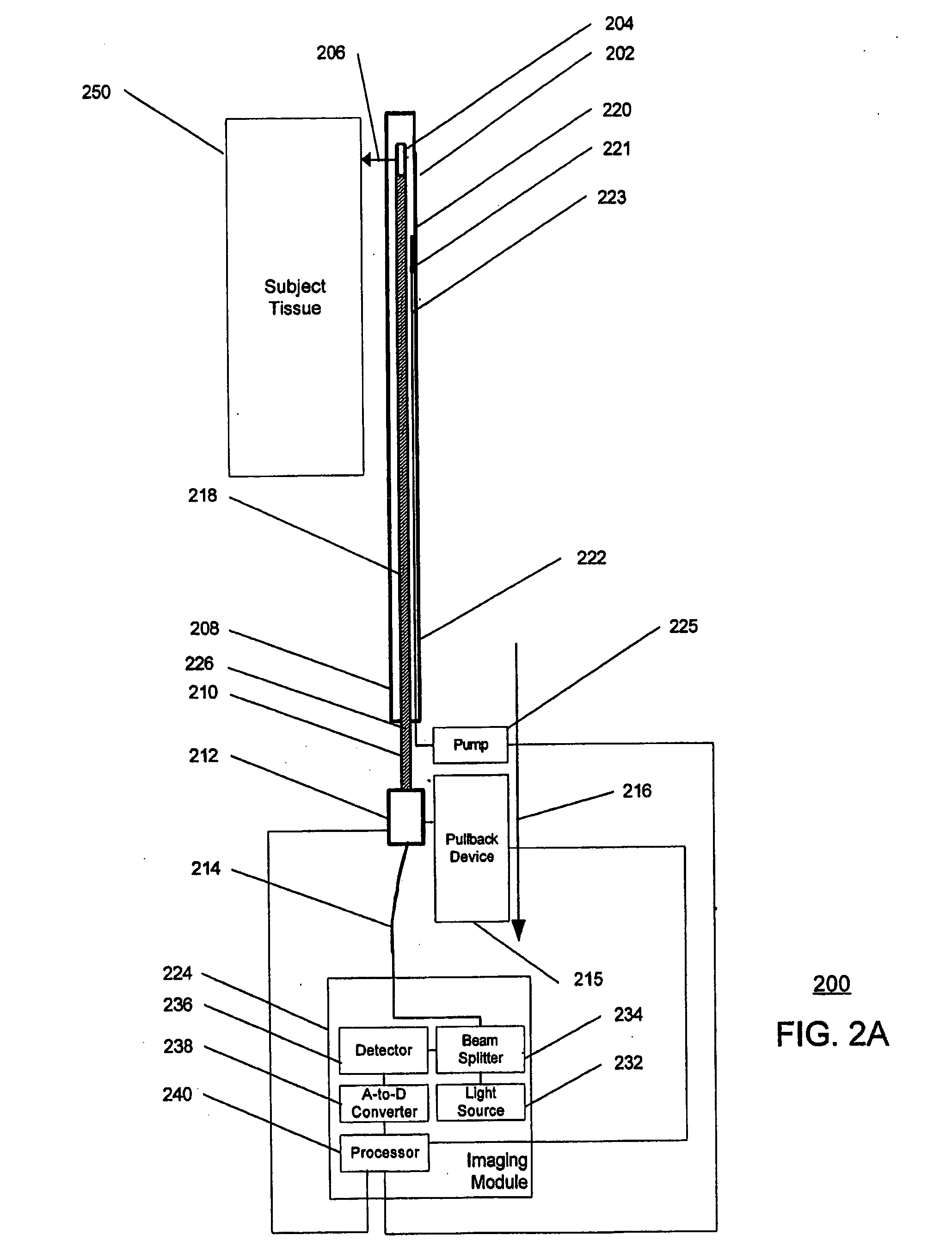
[0033]FIGS. 2A, 2B, 4, 5 and 6 illustrate various exemplary embodiments of an apparatus for obtaining an image of internal surfaces of a segment of an anatomic structure and FIG. 3 shows an exemplary embodiment of a method to implant the same. Generally, the exemplary method and apparatus according to the present invention perform a helical scan of the internal surfaces of the segment of the anatomic structure after injecting a bolus of transparent or semi-transparent fluid, so as to obtain an image of the internal surfaces of the segment of the anatomic structure using an imaging modality. Such technique combines the efficacy of the imaging modality and the process of injecting a bolus of transparent or semi-transparent fluid with the beneficial effect of imaging an entire segment of the anatomic structure. The exemplary embodiments of the method and apparatus according to the present invention utilize a further paradigm for imaging that provide a significant increase in the image ...


PUM


Abstract

An apparatus, method and software arrangement for imaging a surface of a structure that is in contact with an opaque fluid is provided. The apparatus includes an article of manufacture (e.g., a housing), a fluid delivery arrangement and an imaging arrangement. The housing includes an aperture formed in the article of manufacture. The fluid delivery arrangement is configured to deliver a volume of substantially transparent fluid to the aperture formed in the housing. The imaging arrangement is configured to image the surface of the structure using an imaging modality after the volume of the transparent fluid is delivered to the aperture, wherein the imaging arrangement and/or the article of manufacture is translated along the surface of the structure while imaging the surface of the structure.

Description

CROSS-REFERENCE TO RELATED APPLICATION(S) [0001] The present invention claims priority from U.S. Patent Application Ser. No. 60 / 604,138 filed on Aug. 24, 2004, the entire disclosure of which incorporated herein by reference.FIELD OF THE INVENTION [0002] The present invention relates to methods and apparatus that use optical radiation for imaging surfaces and, more particularly to a method and apparatus that use optical radiation to image an interior target surface of a blood vessel. BACKGROUND INFORMATION [0003] Acute myocardial infarction (“AMI”) is the leading cause of death in the United States and industrialized countries. Research conducted for over the past 15 years has demonstrated that several types of minimally or modestly stenotic atherosclerotic plaques, termed vulnerable plaques, are precursors to coronary thrombosis, myocardial ischemia, and sudden cardiac death. Postmortem studies have identified one type of vulnerable plaque, i.e., the thin-cap fibroatheroma (“TCFA”),...

Claims


Application Information

Patent Timeline
Patent Type & Authority Applications(United States)
IPC IPC(8): A61B5/05
CPCA61B5/0062A61B5/0066A61B5/6852A61B5/02007A61B5/0084A61B5/00
Inventor TEARNEY, GUILLERMO J.SHISHKOV, MILENBOUMA, BRETT EUGENE
Owner THE GENERAL HOSPITAL CORP
Why Eureka
Try Eureka