Kit for image guided surgical procedures

a surgical procedure and image technology, applied in the field of image guided surgery, can solve the problems of know, difficult to keep track of the needle tip relative to the patient, drawbacks and limitations of ct fluoroscopy, etc., to reduce the likelihood of trauma or injury, reduce the contamination of the operator's hand, and simple screw system

Inactive Publication Date: 2006-04-13
MURPHY KIERAN P +1
View PDF99 Cites 9 Cited by
  • Summary
  • Abstract
  • Description
  • Claims
  • Application Information

AI Technical Summary

Benefits of technology

[0011] In another aspect of the invention there is provided a biopsy needle wherein the stylet is attached to the trocar via a locking means or attachment means, such as a Luer lock.™ or a simple screw system. The locking biopsy needle is thus used under CT image guidance, advanced using the needle holder. The locking needle thus can be unlocked at the desired time and reduce the likelihood of trauma or injury to the patient during navigation under CT image guidance.
[0012] In another aspect of the invention there is provided a drape that reduces contamination of the operator's hand against the side of the CT scanner. For conventional angiography, a sterilized plastic bag with an elasticated top is placed around the image intensifier and used like a sack. In a CT machine, there is provided a donut-shaped configuration and the patient passes through the central hole of the donut. Preferably, such a drape is disposable, but re-sterilizable drapes are also within the scope of the invention. It is presently preferred that the drape would be like a basketball hoop. In this particular implementation of this aspect of the invention, the basketball-hoop like drape is attachable to the open ends of the CT scanner by any suitable attachment means, such as either or a combination of a) adhesive, b) preplaced hoops affixed to the CT scanner and whereby such hoops would attach by an elasticated band to the drape; c) the drape could be made from a metal that is foldable and therefore transportable, though when released from its package would have a radial force such that it would affix the drape to either side of the CT scanner. Such a material could be Nitinol, from Nitrol Devices and Components, 47533 Westinghouse Drive, Fremont, Calif. 94539.
[0013] In another aspect of the invention there is provided a kit for use in CT guided image fluoroscopy, comprising: (1) a needle holder for keeping the operator's hand out of the beam; (2) a needle of diminished beam hardening artifact inducing potential; (3) a lock to fix the stylet with regard to the trocar in an appropriate position; and (4) a drape to protect the operator's hand from contamination.
[0014] In another aspect of the invention there is provided a kit of parts for use in an image guided surgical procedure using a substantially real time imaging machine comprising: a needle holder having a grasping means and a handle depending therefrom, the handle being configured such that the grasping means can be exposed to the imaging beam and an operator's hand can be distal from the imaging beam in relation to the grasping means; a needle attachable to the grasping means and having a rigidity to travel through mammalian tissue to a target area and having a radioopacity that substantially preserves an appearance of the needle when the needle is viewed on a display of the real time imaging machine; and a sleeve for attachment to the real time imaging machine that provides a substantially sterile operating environment for using the needle when attached to to the machine.
[0016] In another aspect of the invention there is provided a surgical instrument for use in an image guided surgical procedure using a substantially real time imaging machine comprising: a needle holder having a grasping means and a handle depending therefrom, the handle being configured such that the grasping means can be exposed to the imaging beam and an operator's hand can be kept a distance away from the imaging beam; and a needle attachable to the grasping means and having a rigidity to travel through mammalian tissue to a target area and having a radioopacity that substantially preserves an appearance of the needle when the needle is viewed on a display of the real time imaging machine.
[0025] In another aspect of the invention there is provided an imaging machine comprising a channel for receiving a patient and exposing the patient to a substantially real time imaging beam. The machine also includes an attachment means for affixing a sterile drape to the channel, such that when the sterile drape is attached thereto a substantially sterile barrier between the channel and the patient is provided, thereby providing a substantially sterile environment for the patient.

Problems solved by technology

Many of these ultrasound biopsies are performed because computerized tomography (“CT”)is not available.
With this process a biopsy could take hours and it was hard to keep track of the needle tip relative to the patient and know if it was necessary to angle up or down to get to the target.
However, there are drawbacks and limitations to CT fluoroscopy.
This can result in an accumulation of excessive radiation dose to the physician's hand.
The physician may perform the procedure repeatedly during his career or even during a single day and this cumulative dose becomes an issue of personal radiation safety.
Furthermore, current biopsy needles are composed of metal that generates significant artifacts when used with x-ray detectors of CT quality.
These artifacts can obscure the intended target or obscure an important structure and possibly make it possible for inadvertent injury of the target.
Accordingly, current biopsy needles are not generally suitable for CT image guided surgical procedures.
A further disadvantage of the prior art is that needles that are currently used for biopsies typically have the stylet attached to the trocar loosely, yet such a loose attachment can present certain hazards when using such a needle under CT imaging.
A further disadvantage of the prior art is that, since CT machines are typically used for simple capturing of images, they are typically non-sterile, and therefore, under CT image guidance procedures, elaborate sterilization can be necessary to reduce risk of patient infection.

Method used


Image

  • Kit for image guided surgical procedures
  • Kit for image guided surgical procedures
  • Kit for image guided surgical procedures

Examples


Embodiment Construction

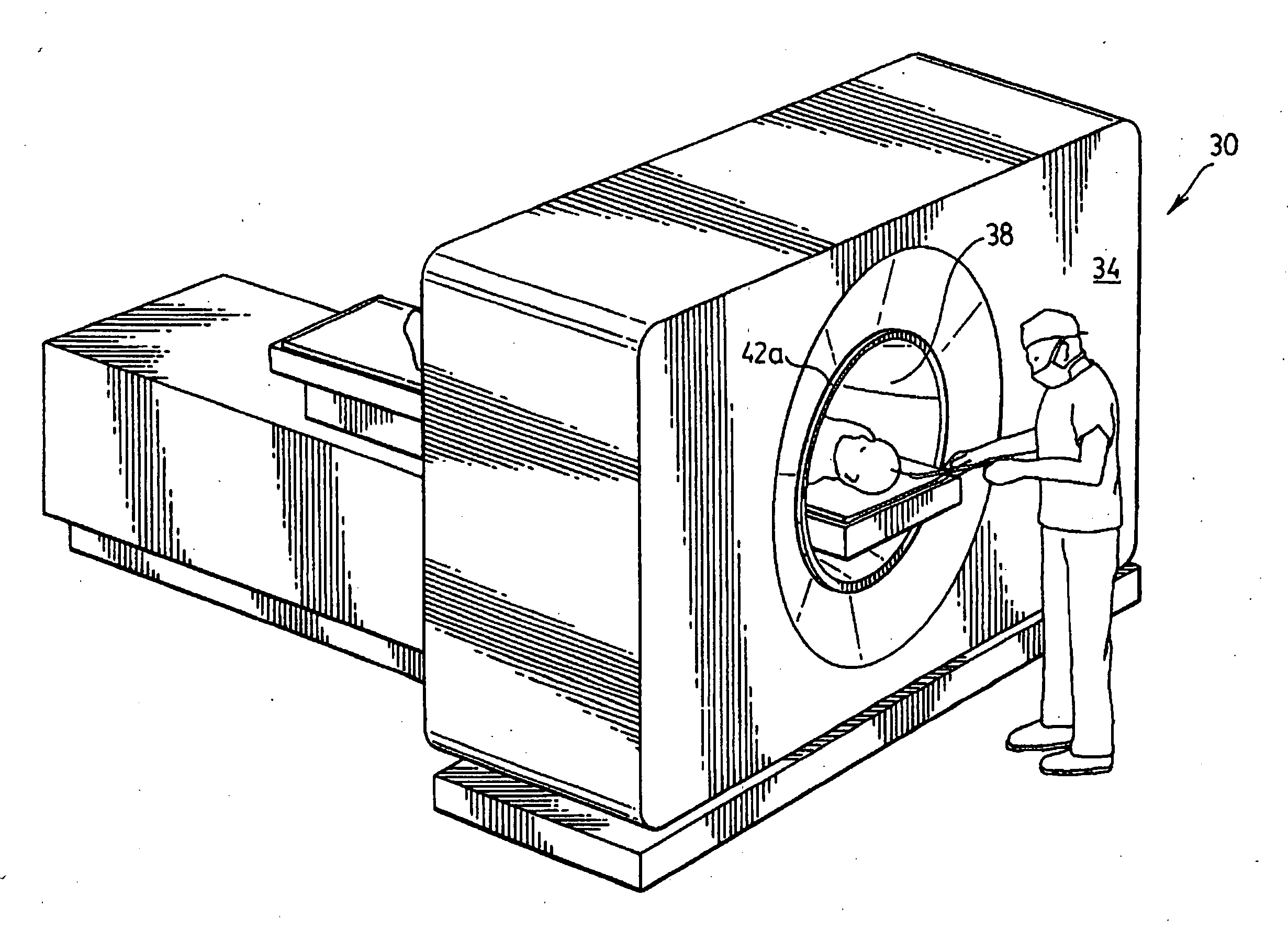
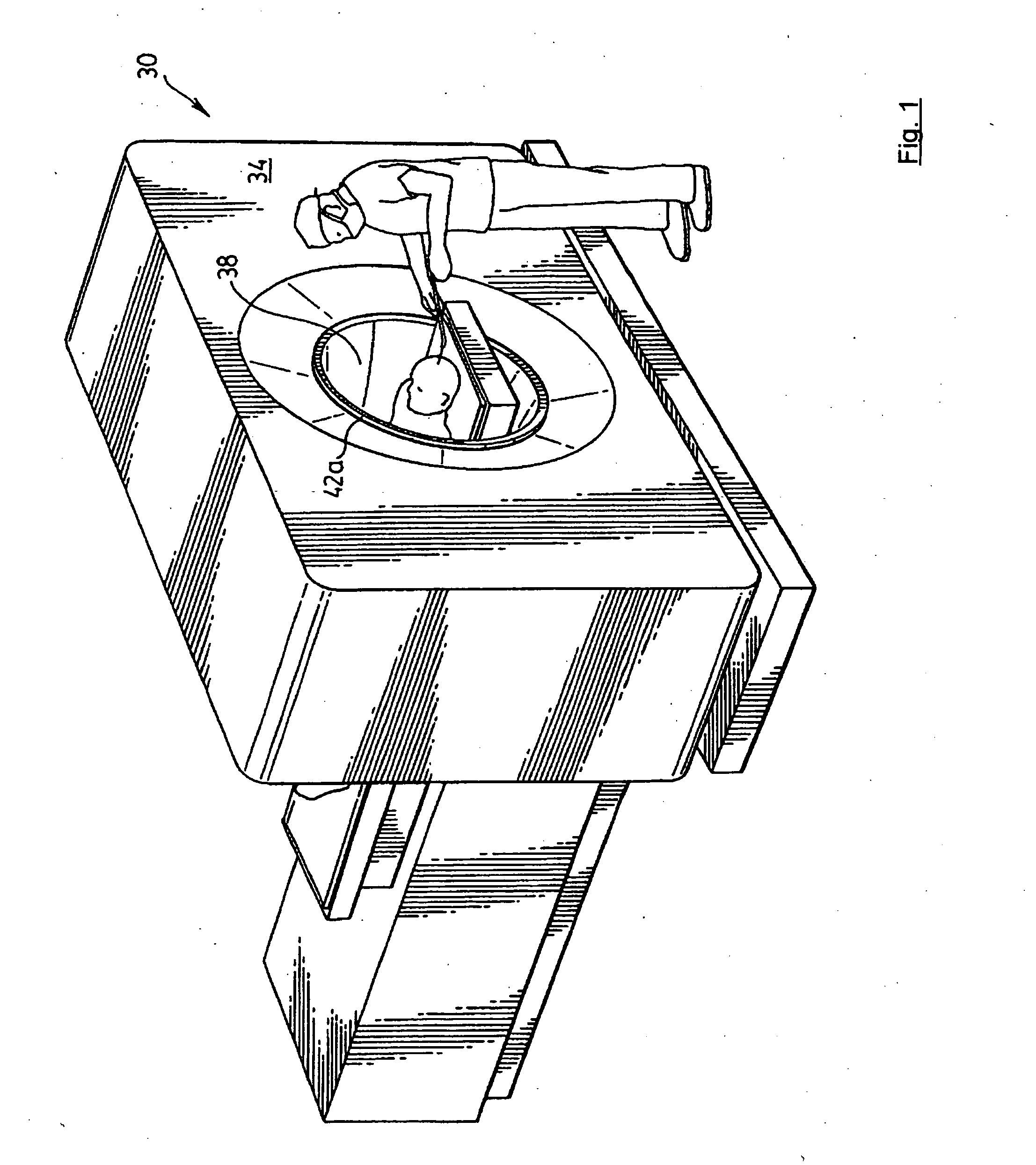
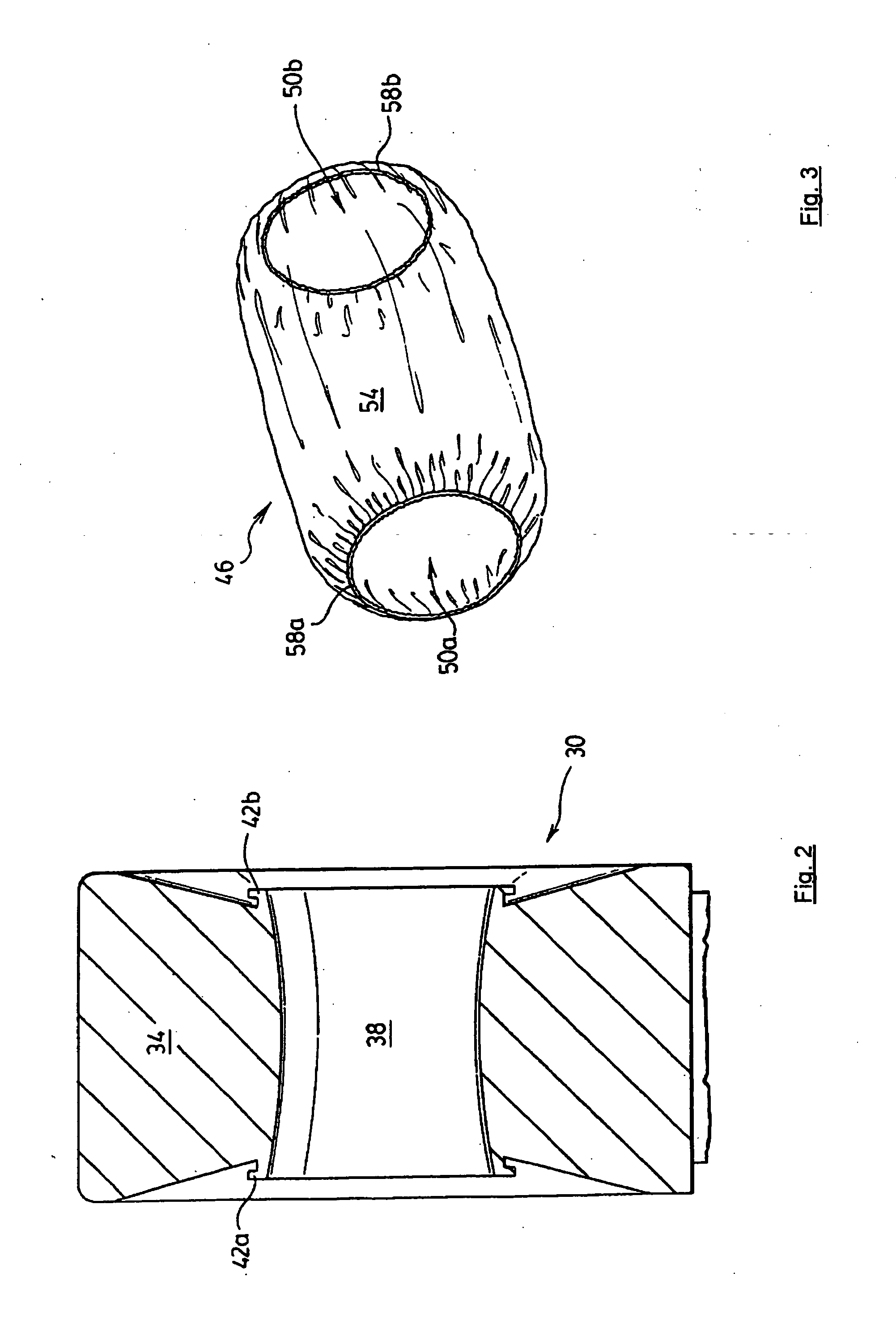
[0040] Referring now to FIGS. 1 and 2, a computerized tomography (“CT”) imaging machine in accordance with an embodiment of the invention is indicated generally at 30. CT Machine 30 is composed of a chassis 34 and a channel 38 through which a patient is received in order to capture the desired images of the patient and / or perform any desired procedures. A presently preferred CT machine for use in the present embodiment is an imaging machine capable of generating substantially real time images. In order to generate images in substantially real time, the imaging machine can generate images at a rate of about fifty frames per second or greater. However, substantially real time images suitable for the present embodiment can also be generated by machines capable of generating images at a rate of about thirty frames per second or greater. However, substantially real time images suitable for the present embodiment can also be generated by a machine capable of generating images at a rate of...


PUM

PropertyMeasurementUnit
angleaaaaaaaaaa
CTaaaaaaaaaa
MRIaaaaaaaaaa

Abstract

An embodiment of the present invention provides a kit of parts for use in a surgical procedure performed under image guidance, and particularly under real time image guidance. The kit includes a sterilized drape for use with the chosen imaging machine and which can be used to provide a sterile operating environment when the procedure is performed under the imaging beam. The kit also includes a needle holder that can keep the surgeon's hand away from the imaging beam. The needle holder is operable to hold a needle that is made from a material suitable for piercing tissue, but also substantially preserves the appearance of the needle when it is viewed under the imaging beam.

Description

PRIORITY CLAIM [0001] The present application is a divisional of U.S. Non-Provisional patent application Ser. No. 10 / 373,835 filed Feb. 27, 2003, which claims priority from U.S. Provisional Patent Application No. 60 / 366,530 filed Mar. 25, 2002 and U.S. Provisional Patent Application No. 60 / 366,529 filed Mar. 25, 2002, and for which co-inventor Murphy of the present application is also named as co-inventor. The contents of all of these documents are incorporated herein by reference.FIELD OF THE INVENTION [0002] The present invention relates generally to image guided surgery and more particularly relates to a kit of parts, and the individual parts of the kit, for use in navigation during a surgical procedure. BACKGROUND OF THE INVENTION [0003] Over one million CT-guided biopsies are performed per year in the US. There are two million ultrasound-guided biopsies a year. Many of these ultrasound biopsies are performed because computerized tomography (“CT”)is not available. Ultrasound is ...

Claims


Application Information

Patent Timeline
Patent Type & Authority Applications(United States)
IPC IPC(8): A61B5/05A61B10/00A61B10/02A61B17/12A61B17/34A61B19/00A61B19/08A61F2/86A61L31/18
CPCA61B6/03A61B10/0233A61B17/3403A61B19/081A61B19/54Y10T428/24273A61B2019/5238A61B2019/5454A61L31/18A61B6/4423A61B2019/5236A61B2090/376A61B46/10A61B2090/374A61B90/39A61B2090/3954
Inventor MURPHY, KIERAN P.GAILLOUD, PHILLIPPE EDMOND
Owner MURPHY KIERAN P
Why Eureka
Try Eureka