Brain function decoding process and system

a brain function and decoding technology, applied in the field of neuroimaging data analysis, can solve the problems of limited resolution, inability to understand how individual brain imaging localizes below the general brain region area of activation, and cannot be applied in medical science, diagnostics, and psychotechnic devices. to achieve the effect of reading the neurologic activity of an individual

Inactive Publication Date: 2006-04-20
MARKS DONALD H
View PDF7 Cites 45 Cited by
  • Summary
  • Abstract
  • Description
  • Claims
  • Application Information

AI Technical Summary

Benefits of technology

[0033] Thus, there exists a need to translate neurologic changes occurring upon sensory or physical stimulation into a decipherable code allowing one to effectively read neurologic activity of an individual.

Problems solved by technology

There has not been an attempt to understand how individual brain imaging is directly linked to the concepts that form the basis of thought.
The data they review, while impressive, does not localize below general brain region areas of activation.
Like all studies to date, they were able to determine gross brain areas of activation, but were limited on their resolution.
At first, the program had trouble matching the monkeys' intentions to the icon's position much more often than chance.

Method used


Image

  • Brain function decoding process and system
  • Brain function decoding process and system
  • Brain function decoding process and system

Examples


example 1

Cognitive Engrams Extracted from Existing Studies

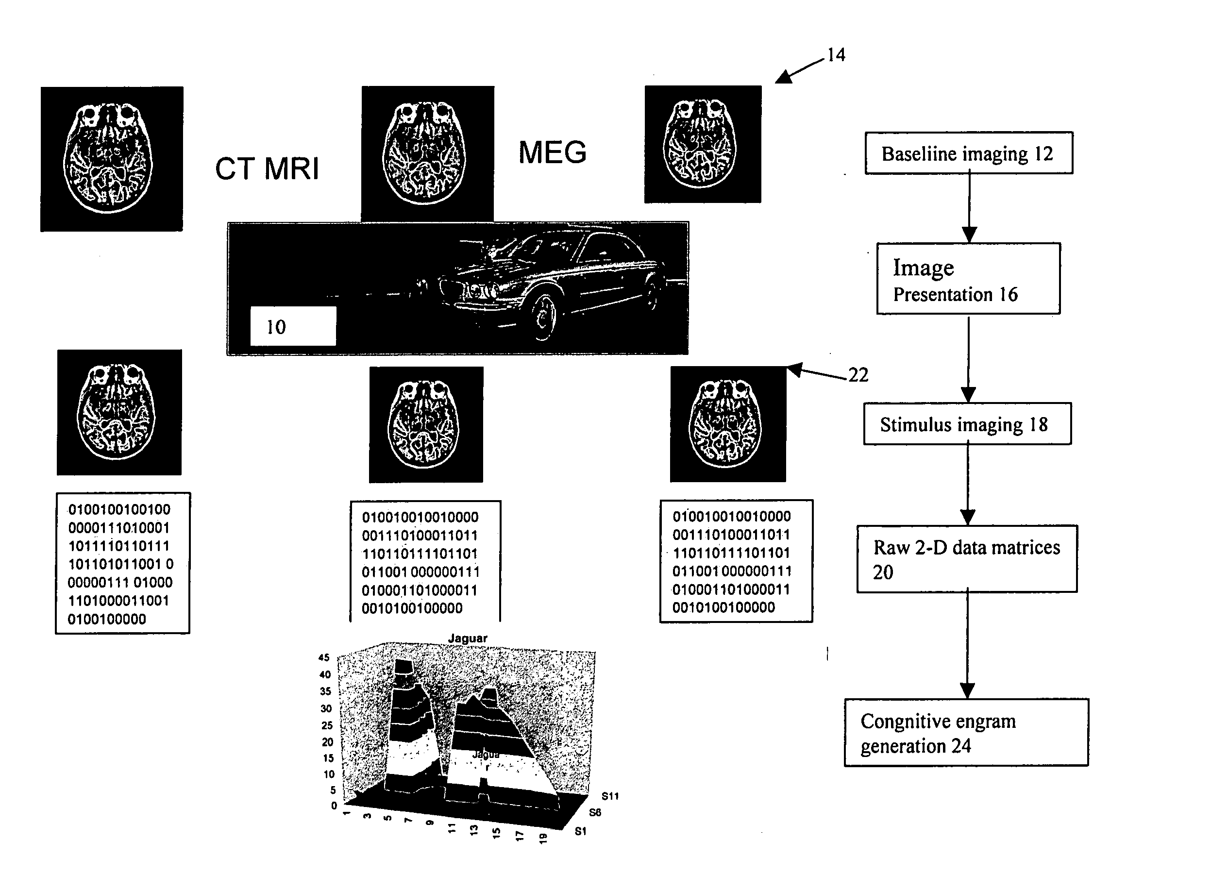
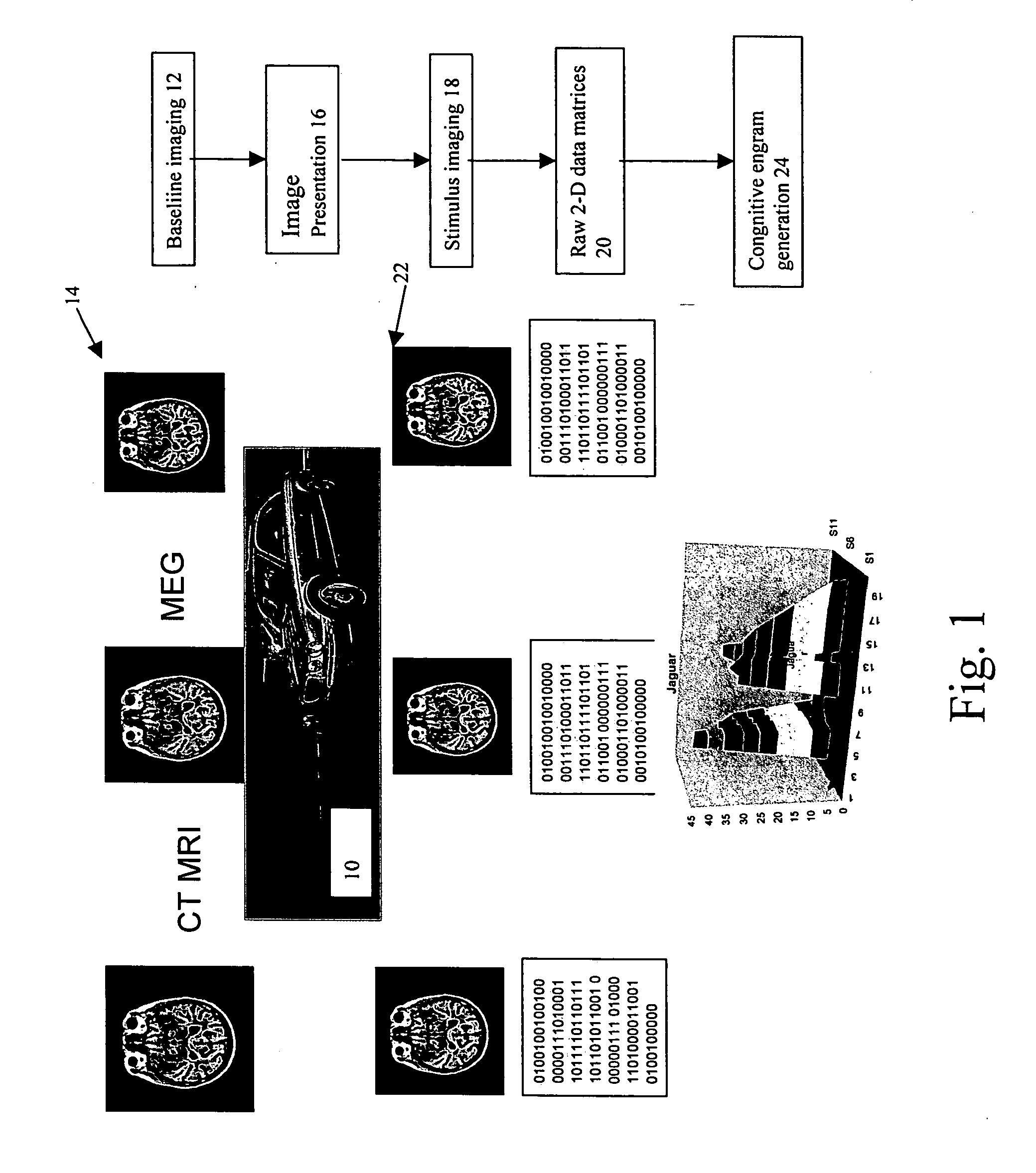
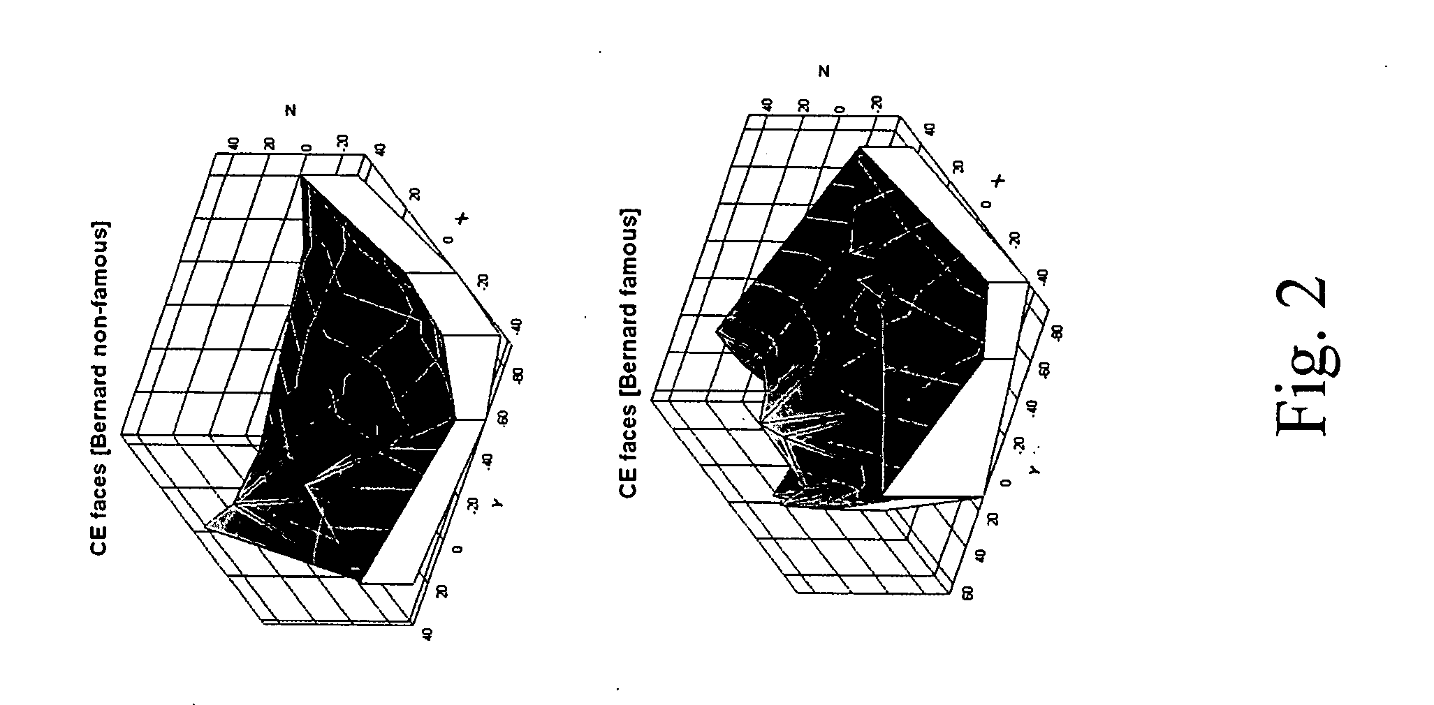
[0059] Recent scientific publications utilizing fMRI to study neurocognitive functions were located by searching the National Library of Medicine. Publications which detailed the specific coordinates of activation areas for visual objects were selected, and are described below. Three-dimensional graphical surface and bubble point representations of the activation maps were constructed by plotting the xyz coordinates. The xyz coordinates were supplied by all three sets of investigators using the normalized space of the Talairach and Tournoux brain atlas (1988).

[0060] All of the publications whose data was reprocessed presented test objects to test subjects while lying in an MRI scanner, using a flat panel display situated to allow visibility during scanning.

[0061] Bernard et al. (2004) studied the presentation and processing of visual imaging of famous and non-famous faces. The study used an event-related fMRI design, utilized a 4-T...

example 2

Distinguishing Cognitive Engrams of Standard Faces

[0064] A 3-T General Electric Signa Excite scanner with whole head coil is used. Changes in the blood oxygen level dependent T2*-weighted MRI signal are measured using a gradient-echo echoplanar sequence. In each time series, 18 (128×128) or 28 (64×64) contiguous axial slices are obtained. There are three sets of scans performed per experiment: localizer, MRI for functional data, and high resolution anatomic.

[0065] The subject lies in the MRI scanner, wearing a phased array MRI head coil as part of the regular head scan. Mounted on the head coil is a 45 degree mirror, allowing them to see down toward their feet and view the test images. Displayed near the bottom of the subject's feet is a large photo, or a white blank paper board, which can have a large X marked across it, which is totally within the field of view of the test subject lying in the scanner. Other means of presentation of visual test images include a goggle image proj...

example 3

Sample Interrogation Protocol

[0077] Background information is obtained on the subject's name, age, sex, SSN, birth city, medications, current and past illnesses, psych history, criminal history, accusation. Then, a basic set of standard images is presented to the test subject while undergoing neuroimaging, in order to obtain a background pattern of activation. Individual responses to questions are recorded via a number of techniques, including verbal and pressing of buttons on handgrips. The test subject is presented with between ten to twenty test questions, and asked to supply intentionally deceptive and intentionally truthful responses to each question.

[0078] The test subject is presented with case-specific questions, with accompanying images / displays of words and phrases concerning: [0079] Facts about their own personal life, name, sex, age, SSN, residence, criminal history; and [0080] Specific photos / questions / statements relating to the subject at issue, including potentially...


PUM


Abstract

A method of interpreting cognitive response to a stimulus is disclosed. The method includes collecting baseline neural activity data from a subject absent a stimulus. Neural activity data is collected while the subject is being stimulated through exposure to a stimulus. A unique three-dimensional cognitive engram is then plotted representative of cerebral regions of stimulated neural activity caused by the stimulus. A novel graphical representation is plotted in three dimensions to indicate the brain region response unique to that stimulus.

Description

RELATED APPLICATION [0001] This application claims priority of U.S. Provisional Patent Application Ser. No. 60 / 620,507 filed Oct. 20, 2004, and U.S. Provisional Patent Application Ser. No. 60 / 637,628 filed Dec. 17, 2004, which are incorporated herein by reference.FIELD OF THE INVENTION [0002] The present invention in general relates to analysis of neuroimaging data and in particular to identification of an individual's response to a sensory or physiologic stimulation through analysis of neuroimaging data. BACKGROUND OF THE INVENTION [0003] Scientific research has demonstrated that memory and consciousness reside in the organization of neurons, their interconnections, charges on the cell surfaces, intracellular and extracellular proteins and other molecules, and other factors. Although the ability to understand the molecular and cellular mechanism of memory and consciousness, the sequences, the charges, and how they relate to memory and consciousness is in very early development, the...

Claims


Application Information

Patent Timeline
Patent Type & Authority Applications(United States)
IPC IPC(8): A61B5/05A61B5/04
CPCA61B5/055A61B5/16
Inventor MARKS, DONALD H.
Owner MARKS DONALD H
Why Eureka
Try Eureka