Eureka AIR delivers breakthrough ideas for toughest innovation challenges, trusted by R&D personnel around the world.

Method for detection of micro-metastasis

a micro-metastasis and detection method technology, applied in the field of cancer detection, can solve the problems of complicated routine use, the controversial use of tumor suppressors, and the risk of systemic recurrence for most common types of human cancers

Inactive Publication Date: 2006-06-08
YISSUM RES DEV CO OF THE HEBREWUNIVERSITY OF JERUSALEM LTD +2
View PDF2 Cites 10 Cited by
  • Summary
  • Abstract
  • Description
  • Claims
  • Application Information

AI Technical Summary

Benefits of technology

The present invention is based on the discovery that by detecting the presence of a specific gene called H19 mRNA in a sample containing cells from a cancer patient, it is possible to identify the presence of minute amounts of circulating cancer cells or micro-metastasis in the patient. This is done by detecting the presence of H19 mRNA in a sample containing cells from a cancer patient, and comparing the amounts of H19 mRNA in the sample to a standard calibration curve of H19 mRNA as a function of the number of cancer cells. The invention thus provides a sensitive and reliable method for detecting residual cancer cells or cancer cells from micrometastasis in a patient.

Problems solved by technology

The proposal that H19 is a tumor suppressor has been controversial, however.
However, the tiny size of micro-metastasis and low number of tumor cells, particularly in the circulation and bone marrow, have presented a challenge for their detection in a reliable and sensitive manner.
The most common types of human cancers bear a considerable risk of systemic recurrence even when they diagnosed and despite curative resection of the primary tumor.
Nevertheless, the microscopic, preferably double-blinded examination of cytocentrifuged bone marrow cells is laborious and observer dependent, thus complicating routine use.
However, low-level gene expression in nonmalignant cells appears to limit the specificity of most candidate PCR markers, with only a few exceptions including PSA in prostate cancer.
However, recent studies have shown several of these markers to be expressed in normal cells of peripheral blood, lymph nodes, and / or bone marrow yielding false-positive results.
These findings may contribute to the lack of consistent correlations between any single tumor marker and well-known clinical and pathological prognostic factors.
Currently there is no consensus recommendation for the routine use of molecular markers in monitoring disease detection in blood or other body fluids.

Method used


Image

  • Method for detection of micro-metastasis
  • Method for detection of micro-metastasis
  • Method for detection of micro-metastasis

Examples


example 1

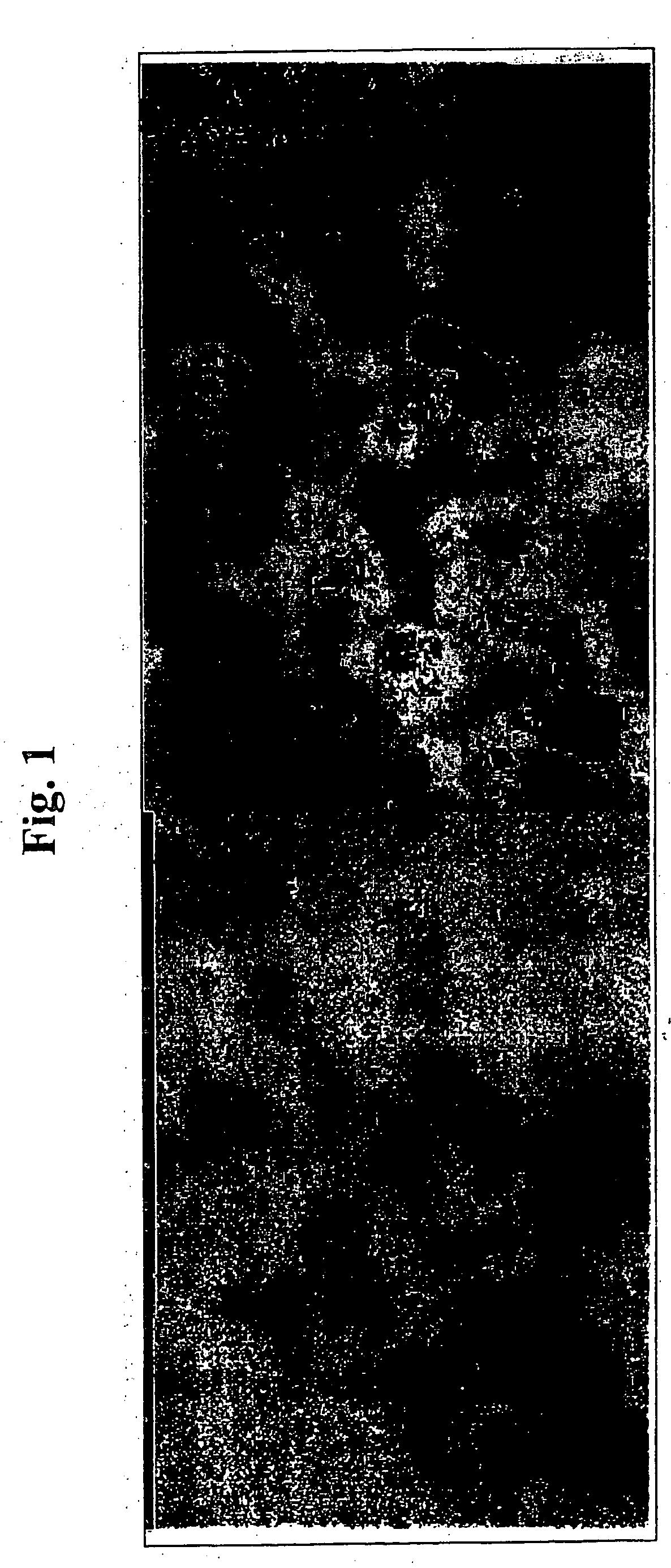
Detection of H19 in Bladder Rinse Fluid

[0097] Voided urine was taken from patient with bladder carcinoma (DIG-H19). The exfoliated cells in the urine were separated from the liquid and underwent in situ hybridization with a radioactive H19 probe as described in I(c) in experimental procedures above.

[0098] The results of the in situ hybridization are shown in FIG. 1. As can be seen the cells present in the urine of cancer patient reacted significantly with the labeled probe, while normal urine (data not shown) did not hybridize with the probe.

example 2

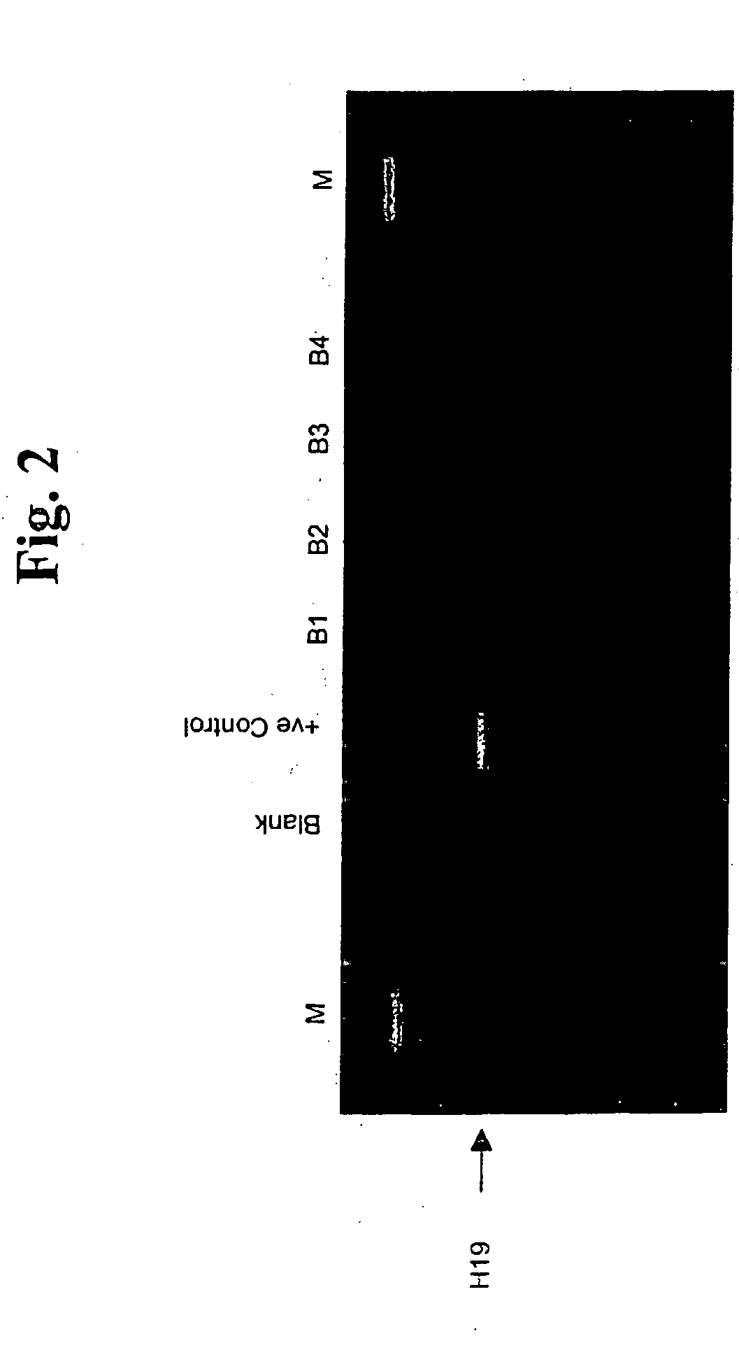
Detection of H19 in Blood Samples of Colon Patients

[0099] Blood from 4 diagnosed colon cancer patients was colleted and prepared as in I(a) above and the H19 mRNA was amplified by RT-PCR as disclosed in I(b) above. The amplification products were separated on a gel and the results are shown in FIG. 2. As can be seen patients B1 and B2 were strongly positive for H19 expression (as compared to blank) while patient B3 showed a week expression of H19, indicating that 3 out of the 4 colon cancer patients had H19 expression in a detectable level.

example 3

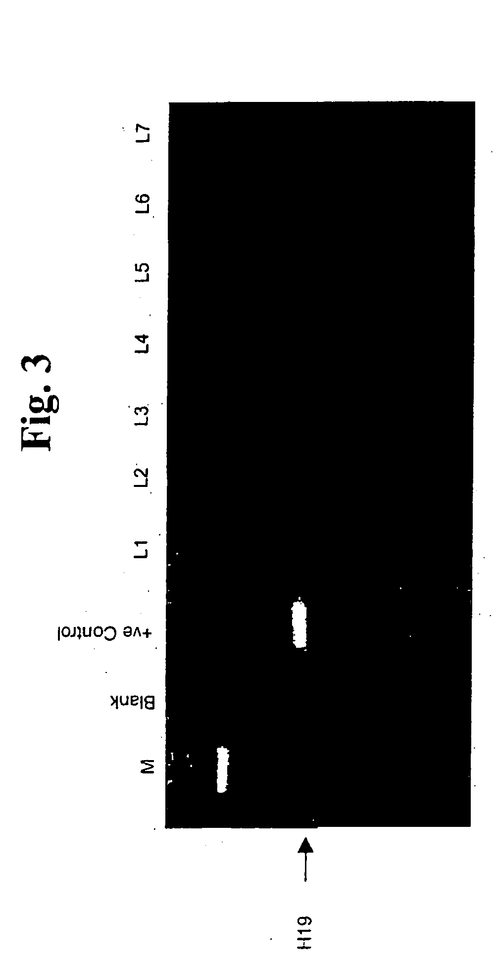
Detection of H19 in Lymph Nodes Obtained from Breast Cancer Patients

[0100] Sentinel lymph nodes were obtained from breast cancer patients as described in I(e) above. The RT-PCR was performed on the extracted mRNA as described in I (b) above and the amplification results were separated on a gel.

[0101] The results are shown in FIG. 3. As can be seen patients L5 and L6 were tested positive for H19 expression indicating that H19 detection can be carried in a lymph node sample.


PUM

PropertyMeasurementUnit
PSAaaaaaaaaaa
threshold RNA levelaaaaaaaaaa
sizeaaaaaaaaaa

Abstract

The present invention concerns a method for the identification of micro-metastasis or residual cancer cells, by identifying the presence of H19 in a sample.

Description

FIELD OF THE INVENTION [0001] The invention is in the field of cancer detection. More specifically, the invention relates to the detection of micro-metastasis. BACKGROUND OF THE INVENTION [0002] The H19 gene is one of the few genes known to be imprinted in humans (Hurst et al., 1996, Nature Genetics 12:234-237). At the very beginning of embryogenesis, H19 is expressed from both chromosomal alleles (DeGroot et al., 1994, Trophoblast 8:285-302). Shortly afterwards, silencing of the paternal allele occurs, and only the maternally inherited allele is transcribed. [0003] H19 is abundantly expressed during embryogenesis, and was first identified as a gene that was coordinately regulated with alpha-fetoprotein in liver by the trans-acting locus raf (Pachnis et al., 1984, Proc. Natl. Acad. Sci. USA 81:5523-5527). Additionally, H19 has been independently cloned by a number of groups using screens aimed at isolating genes expressed during tissue differentiation. For example, Davis et al. (198...

Claims


Application Information

Patent Timeline
Patent Type & Authority Applications(United States)
IPC IPC(8): C12Q1/68
CPCC12Q1/6886C12Q2600/112C12Q2600/158
Inventor HOCHBERG, ABRAHAMAYESH, SUHAIL
Owner YISSUM RES DEV CO OF THE HEBREWUNIVERSITY OF JERUSALEM LTD
Why Eureka
Eureka Blog
Learn More