Live bioload detection using microparticles

a technology of microparticles and bioload, which is applied in the field of live bioload detection using microparticles, can solve the problems of not being able to distinguish quickly and/or easily the presence of live cells from dead cells, and requiring long periods of time, hours or days) before detection, and achieves the effect of rapid detection

Active Publication Date: 2012-01-12
3M INNOVATIVE PROPERTIES CO
View PDF0 Cites 66 Cited by
  • Summary
  • Abstract
  • Description
  • Claims
  • Application Information

AI Technical Summary

Benefits of technology

[0006]In general, the present disclosure relates to articles and methods for detecting live cells in a sample. The articles and methods make possible the rapid detection (e.g., through fluorescence, chemiluminescence, or a color reaction) of the presence of cells such as bacteria on a surface. In some embodiments, the inventive articles are “sample-ready”, i.e., the articles contain all of the necessary features to detect living cells in a sample. In some aspects, the inventive articles and methods provide a means to distinguish a biological analyte, such as ATP or an enzyme, that is associated with eukaryotic cells (e.g., plant or animal cells) from a similar or identical biological analyte associated with prokaryotic cells (e.g., bacterial cells). Furthermore, the inventive articles and methods provide a means to distinguish a biological analyte that is free in the environment (i.e., an acellular biological analyte) from a similar or identical biological analyte associated with a living cell.
[0007]Methods of the present disclosure allow an operator to concentrate cells from a liquid sample and to detect an analyte associated with the cells. In certain embodiments, detection of the analyte may be an indicator of live cells including, in particular, live microbial cells in the sample. In some embodiments, the methods provide for the operator to measure the amount of a biological analyte in the sample. In some embodiments, the methods provide for the operator to, after a predetermined period of time during which an effective amount of a cell extractant is released from a composition into the liquid mixture, measure the amount of a biological analyte to determine differentially the amount of biological analyte from acellular material and from live cells in the sample. In some embodiments, the methods provide for the operator, within a first predetermined period of time, to perform a first measurement of the amount of a biological analyte and, within a second predetermined period of time during which an effective amount of cell extractant is released from the composition, perform a second measurement of the amount of biological analyte to detect the presence of live cells in the sample. In some embodiments, the methods can allow the operator to distinguish whether biological analyte in the sample was released from live plant or animal cells or whether it was released from live microbial cells (e.g., bacteria). The present invention is capable of use by operators under the relatively harsh field environment of institutional food preparation services, health care environments and the like.

Problems solved by technology

Although the above tests are useful in the detection of a contaminated surface, they can require numerous steps to perform the test, they may not be able to distinguish quickly and / or easily the presence of live cells from dead cells and, in some cases, they can require long periods of time (e.g., hours or days) before the results can be determined.
The presence of extracellular material (e.g., non-cellular ATP released into the environment from dead or stressed animal cells, plant cells, and / or microorganisms) can create a high “background” level of ATP that can complicate the detection of live cells.

Method used


Image

  • Live bioload detection using microparticles
  • Live bioload detection using microparticles
  • Live bioload detection using microparticles

Examples


example 1

Incorporation of Cell Extractant into Hydrogel Beads after Polymerization of the Hydrogel

[0202]Hydrogel beads were prepared as described in example 1 of the International Patent Publication No. WO 2007 / 146722. Active beads were prepared by drying as described in example 19 and then soaking in active solution as described in example 23 of the International Patent Publication No. WO 2007 / 146722. One gram of beads was dried at 60° C. for 2 h to remove water from the beads. The dried beads were soaked in 2 grams of 50% (w / v) aqueous solutions of BARDAC 205M (Lonza Group Ltd., Valais, Switzerland) for at least 3 hrs to overnight at room temperature. After soaking, the beads were poured into a Buchner funnel to drain the beads and then rinsed with 10 to 20 ml of distilled water. The excess water was removed from the surface of the beads by blotting them with a paper towel. The beads were stored in a jar at room temperature for at least two weeks before they were used.

example 2

Cell Concentration by Use of Microparticles and Detection Using Cell Extractant-Loaded Hydrogels and ATP Bioluminescence

[0203]3M™ CLEAN-TRACE Surface ATP system was obtained from 3M Company (St. Paul, Minn.). Pure cultures of E. coli ATCC 51183 were inoculated into tryptic soy broth and grown overnight at 37° C. The bacterial culture was diluted to approximately 106 or 105CFU / ml in Butterfield's buffer (pH 7.2±0.2; monobasic potassium phosphate buffer solution; VWR, West Chester, Pa.) and 100 microliters of the diluted suspension were added directly to individual tubes containing ten ml of deionized water (Milli-Q Biocel System, Millipore, MA) samples to obtain approximately 105 CFU or 104 CFU in ten ml, respectively. Ten mg of autoclaved CM-111 3M™ Cosmetic Microspheres (calcined amorphous spheroidized magnesium silicate powders; 3M Company; St. Paul, Minn.) were added to the tubes containing cells and mixed at room temperature for about 15 min. The particles were allowed to settle...

example 3

Detection of Microbial Cells in a Unitary Sample Preparation and Detection Device Using an ATP Bioluminescence Detection System

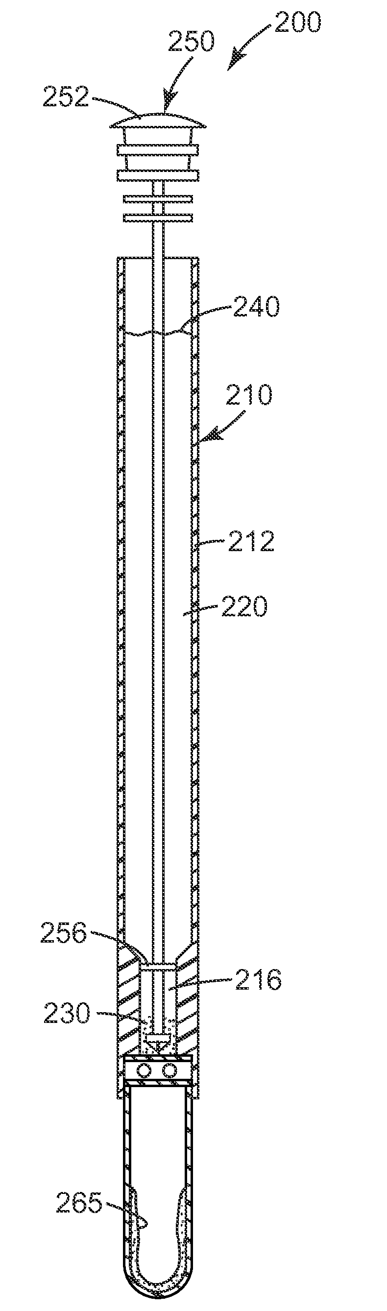
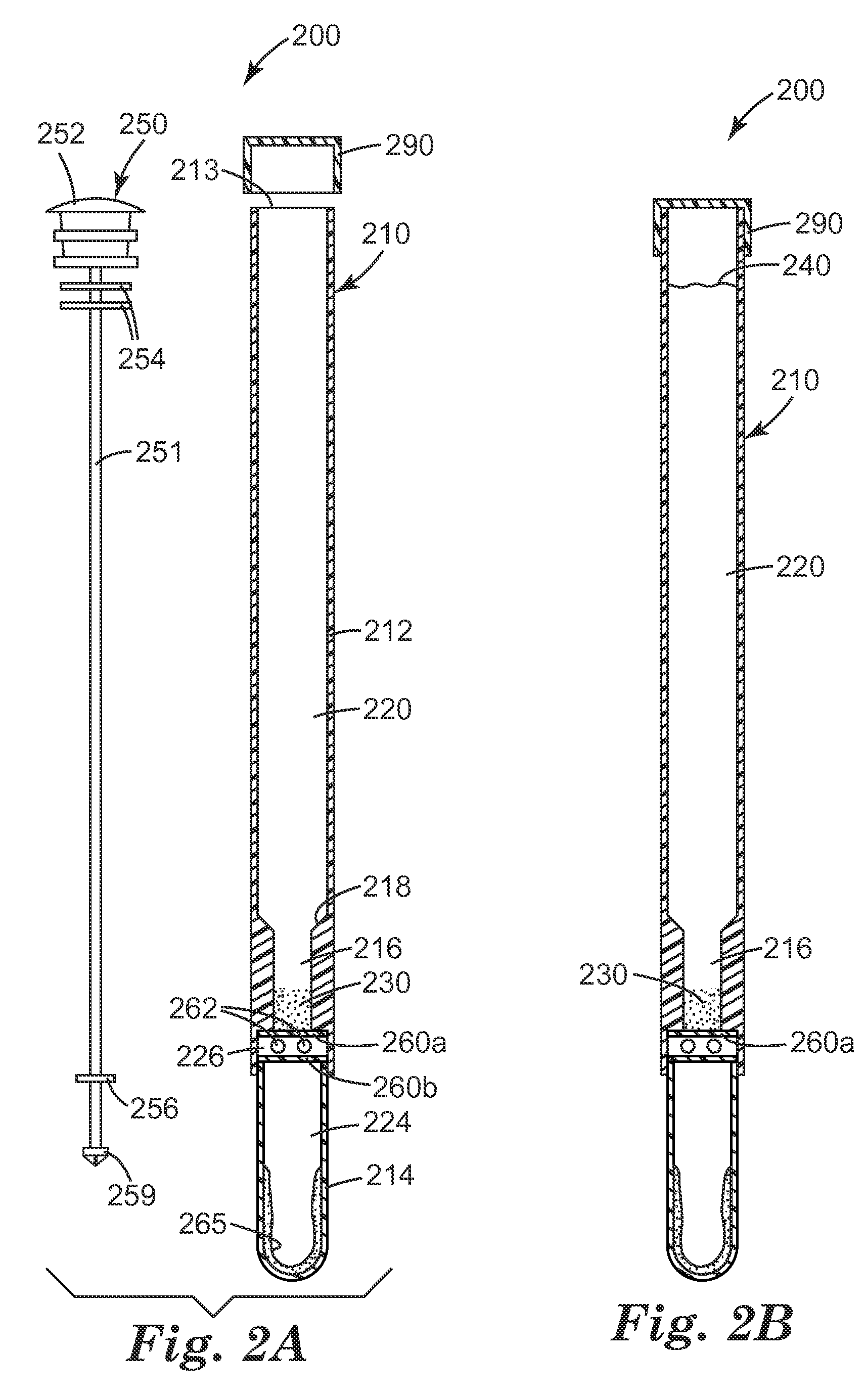
[0205]A unitary sample preparation and detection device 200, as shown in FIG. 2, is used in this Example. The device contains approximately 10 mg of autoclaved CM-111 3M Cosmetic Microspheres in the upper receptacle 220. Lower receptacle 224 contains a liquid detection reagent 265, which consists of approximately 0.6 milliliters of the luciferase / luciferin liquid reagent solution from a CLEAN-TRACE surface ATP system. The third receptacle 226 contains two BARDAC 205M beads made according to Preparative Example 5 of U.S. Patent Application No. 61 / 101,546, filed Sep. 30, 2008. Ten milliliters of sterile deionized water is added to the upper receptacle 220 of the unitary devices 200 immediately before use.

[0206]E. coli overnight cultures are prepared as described in Example 2. The bacterial culture is diluted to approximately 106 or 105 CFU / ml in Butterfield's ...


PUM

PropertyMeasurementUnit
particle sizesaaaaaaaaaa
sizeaaaaaaaaaa

Abstract

The present invention provides methods to concentrate cells onto microparticles, to concentrate the microparticles, and to detect the cells. The present invention also includes unitary sample preparation and detection devices to be used in accordance with the methods.

Description

CROSS REFERENCE TO RELATED APPLICATIONS[0001]This application claims the benefit of U.S. Provisional Patent Application No. 61 / 141,685, filed Dec. 31, 2008 and U.S. Provisional Patent Application No. 61 / 291,301, filed Dec. 30, 2009, which are incorporated herein by reference.BACKGROUND[0002]Various tests are available that can be used to assess the presence of biological analytes in a sample (e.g. surface, water, air, etc). Such tests include those based on the detection of ATP using the firefly luciferase reaction, tests based on the detection of protein using colorimetry, tests based on the detection of microorganisms using microbiological culture techniques, and tests based on detection of microorganisms using immunochemical techniques. Surfaces can be sampled using either a swab device or by direct contact with a culture device such as an agar plate. The sample can be analyzed for the presence of live cells and, in particular, live microorganisms.[0003]Results from these tests a...

Claims


Application Information

Patent Timeline
Patent Type & Authority Applications(United States)
IPC IPC(8): C12Q1/66C12M1/34C12Q1/68C12Q1/48C12Q1/04G01N33/569
CPCA61B10/0045A61B10/02B01L3/5082B01L2200/026B01L2200/10C12Q1/04C12M23/32C12M23/34C12M33/02G01N33/56916G01N2333/245C12M23/08B01L3/5021
Inventor RAJAGOPAL, RAJHALVERSON, KURT J.KSHIRSAGAR, MANJIRI T.AYSTA, JAMES E.
Owner 3M INNOVATIVE PROPERTIES CO
Why Eureka
Try Eureka