Methods and probes for detecting esophageal cancer

a technology probes, applied in the field of methods and probes for detecting esophageal cancer, can solve the problems of poor inter-observer reproducibility of pathologists for the diagnosis of lgd and hgd, not frequently followed, limited sampling, etc., and achieve the effect of better results

Active Publication Date: 2006-09-21
ABBOTT MOLECULAR INC +1
View PDF4 Cites 30 Cited by
  • Summary
  • Abstract
  • Description
  • Claims
  • Application Information

AI Technical Summary

Benefits of technology

[0007] Combinations of individual probes within a probe set of the present invention are to be chosen for combined sensitivity and specificity when used in the methods of the present invention. Chromosomal probes that detect the most frequent chromosomal losses or gains associated with an esophageal carcinoma and / or dysplasia are to be chosen, as are probes that complement one another based on sensitivity, specificity, and detectability. In this invention, probe sets chosen for the identification of LGD will have DFI values that are at most about 0.7. Probe sets chosen for the identification of HGD+EA will have DFI values that are at most about 0.5. In either case, DFI values of less than 0.5 usually provide even better results while DFI values of at most about 0.35 usually provide even better results.

Problems solved by technology

However, this recommendation is not frequently followed mainly due to the extended length of time needed to perform this procedure, especially on patients with long segment BE.
Problems associated with the use of biopsies for monitoring Barrett's patients for the development of neoplasia include: 1) limited sampling of affected mucosa, 2) impracticability of taking four-quadrant biopsies every 1-2 cm, and 3) poor inter-observer reproducibility of pathologists for the diagnosis of LGD and HGD.
This limited sampling may lead to false negative pathology results or to under-staging (e.g., pathology results showing only IM or LGD in a patient who has HGD or EA).

Method used


Image

  • Methods and probes for detecting esophageal cancer
  • Methods and probes for detecting esophageal cancer
  • Methods and probes for detecting esophageal cancer

Examples


example 1

Probe Selection

FISH Probe Sets

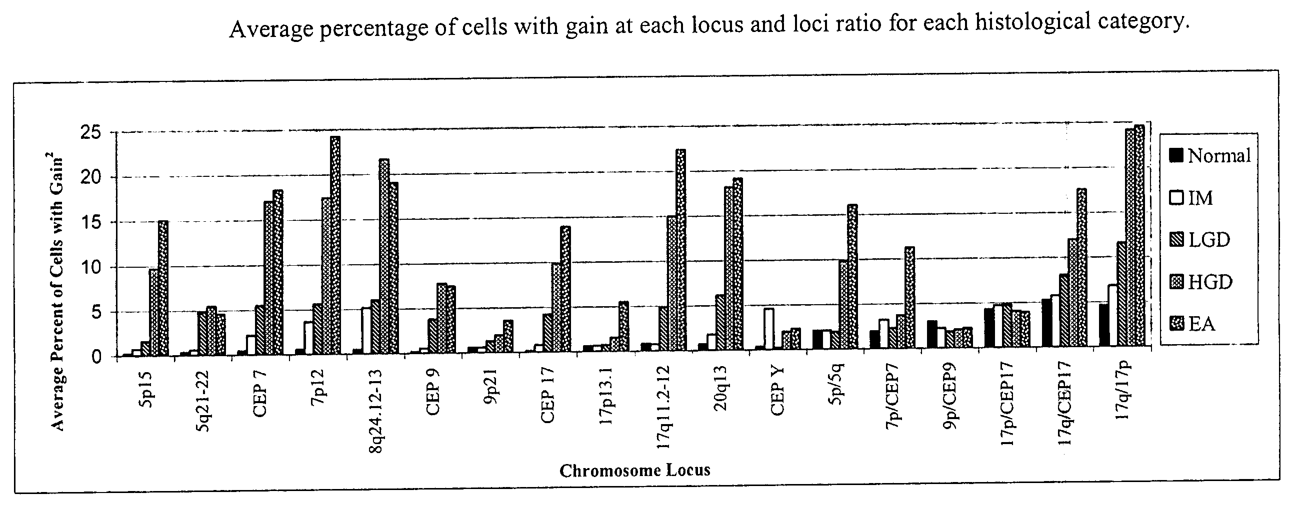
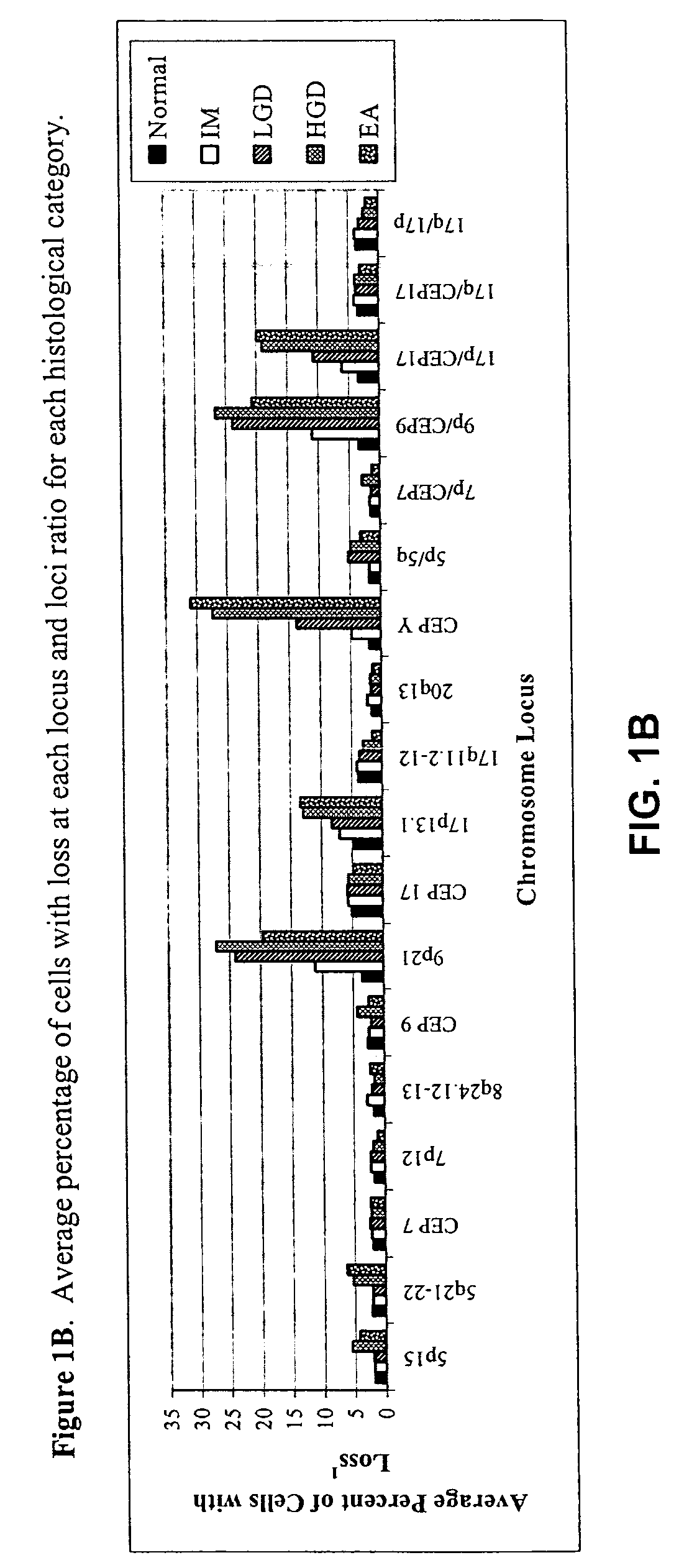
[0057] FISH was performed with three unique probe sets. Each probe set contained four chromosome enumeration probes (CEP®) or locus specific identifiers (LSI®) to centromeres or specific loci of chromosomes that have been shown to be frequently altered in patients with BE-associated neoplasia (Table 1). The CEP 7, CEP 9, and CEP 17 probes were included to determine allelic gain or loss of the corresponding LSI probes on those chromosomes (e.g., 9p21 on chromosome 9) or aneusomy of those chromosomes.

TABLE 1FISH Probes and Gene Target Locations Used for Probe SelectionProbeSetRedGreenAquaGoldILSI 9p21LSI 5p15CEP 9LSI 5q21-22(P16)(APC)IICEP YLSI 17q11.2-12CEP 17LSI 17p13.1 (P53)(HER2 / NEU)IIILSI 20q13.2LSI 8q24.12-13CEP 7LSI 7p12 (EGFR)(C-MYC)

With the exception of the LSI® 5q21-22 (APC) probe, the LSI® and CEP® probes are commercially available from Vysis, Inc. (Downers Grove, Ill., www.vysis.com) labeled with SpectrumOrange™. Instead of the Spectrum...

example 2

Esophageal Cancer Detection

[0092] As a non-limiting exemplification of the present invention, the four-color probe set 8q24.12-13, 9p21, 17q11.2-12 and 20q13, described in Example 1 above, was used to assess esophageal brushing samples for the presence of cells that have chromosomal abnormalities consistent with a diagnosis of LGD, HGD, or EA. Samples were prepared for FISH hybridization and subject to hybridization with the probe set as described in the probe selection study (Example 1, above) and as described below. For cases in which the initial 100-cell enumeration was negative for polysomy, the remainder of the slide was scanned for morphologically abnormal cells (e.g., nuclear enlargement, nuclear irregularity and mottled chromatin staining) and their FISH hybridization patterns of these cells also recorded.

Cell Harvest

[0093] A 50 mL centrifuge and a 1.8 ml micro-centrifuge tube were labeled with appropriate patient identifiers. The specimen container (PreservCyt™ solutio...


PUM

PropertyMeasurementUnit
temperatureaaaaaaaaaa
volume ratioaaaaaaaaaa

Abstract

Probe sets and methods of using probes and probe sets for selectively detecting high grade dysplasia and esophageal adenocarcinoma or low grade dysplasia from biologic samples are described. Methods of the invention include contacting a biological sample obtained from a subject with a set of chromosomal probes to selectively detect an esophageal carcinoma or precursor lesion in the sample, if any, under conditions for specifically hybridizing the probes to their nucleic targets present in the sample. The presence or absence of high grade dysplasia and esophageal adenocarcinoma or low grade dysplasia is thereafter specifically determined from the hybridization pattern detected for the set of chromosomal probes to the biological sample.

Description

BACKGROUND OF THE INVENTION [0001] It is estimated that there will be 14,250 new cases and approximately 13,300 deaths from esophageal cancer in the United States during 2004. Approximately 80% of these tumors will be esophageal adenocarcinoma (EA) and the remaining 20% will be squamous cell carcinoma. The majority, if not all, of EA are thought to arise in patients with Barrett's esophagus (BE), a pre-neoplastic condition caused by metaplasia of the normal squamous mucosa of the distal esophagus into specialized intestinal mucosa containing goblet cells. BE is caused by chronic gastroesophageal reflux disease (GERD), a disorder that affects more than 20 million Americans on a daily basis. Six to fourteen percent of people with chronic GERD will develop BE. The incidence of EA in patients with BE has been reported to be approximately 0.5%-1.0% per year and the lifetime cancer risk for patients with BE is about 5%. [0002] The histologic steps leading to esophageal adenocarcinoma in p...

Claims


Application Information

Patent Timeline
Patent Type & Authority Applications(United States)
IPC IPC(8): C12Q1/68
CPCC12Q2600/112C12Q1/6886
Inventor HALLING, KEVINMORRISON, LARRY E.BRANKLEY, SHANNON
Owner ABBOTT MOLECULAR INC
Why Eureka
Try Eureka